
What are myomas? In Italy National Cancer Institute study uses radiomics to diagnose uterine fibroids
Myomas or uterine fibroids are very common benign and often asymptomatic neoformations resulting from an excessive proliferation of smooth muscle tissue
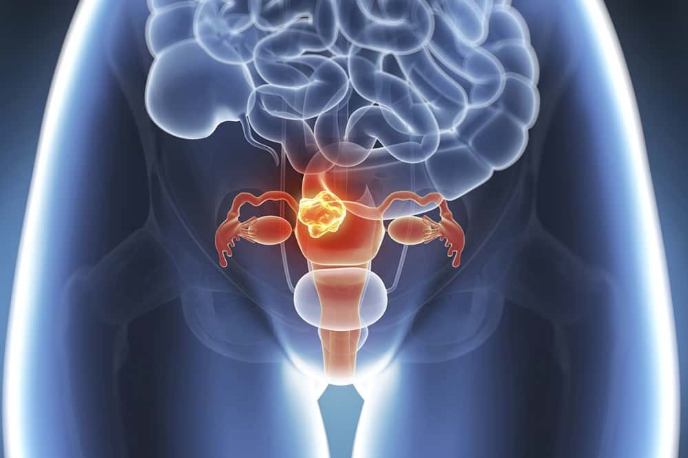
Fibroids usually arise on the uterine wall (myometrium), and more rarely can grow at the ovarian level or separate from the uterus to which they remain connected by a small stalk.
The size of myomas varies greatly from a few mm to over 20 cm
Myomas can be asymptomatic and be diagnosed during a routine check-up, or they can be associated with disabling symptoms including: haemorrhagic menstrual cycles and anaemia, pelvic pain, pain during intercourse (dysmenorrhoea), urinary urgency, infertility, miscarriage and pregnancy complications.
The causes leading to the formation of uterine myomas are not completely known, although a genetic predisposition and subsequent susceptibility to hormonal stimulation have been recognised.
The malignant counterpart of myomas, sarcoma, is a rare and often fatal tumour
The correct diagnosis of myomas and sarcomas is a real challenge for the gynaecologist and ultrasound is the diagnostic method of first choice; ultrasound is always available to the gynaecologist, fast, inexpensive and requires no preparation on the part of the patient.
Unfortunately, ultrasound is operator-dependent and good operator training is essential for an accurate examination.
The treatment of myomas is chosen according to their location, size, age of the patient, her health condition and her desire for offspring.
Surgery for uterine myomas aims to be conservative and minimally invasive, which is why it is often proposed to young patients who have not yet completed their reproductive project.
Unfortunately, to date, there are no diagnostic methods capable of distinguishing with good accuracy malignant neoformations from myomas, with the risk of inadequate surgery in the case of occult sarcoma and a worsening of the patient’s prognosis.
In this regard, in 2014, the US Food and Drug Administration (FDA) warned healthcare professionals against the use of the uterine morcellator, a surgical tool used to ‘fragment’ large myomas (which are then shredded and can be removed through laparoscopic trocars), as it could promote the dissemination of tumour debris in the abdomen in the case of an occult malignant lesion.

An accurate preoperative diagnosis is essential in order to plan an adequate surgery (conservative in case of myoma, demolitive in case of uterine sarcoma).
A study[1] was recently published in Gynecologic Oncology, the result of the work of researchers at the Istituto Nazionale dei Tumori di Milano in collaboration with the university spin-off DeepTrace Technologies IUSS-Pavia, precisely to meet this need.
Myomas: researchers have created a reproducible model using radiomics applied to ultrasound images
The images of uterine neoformations, in this case obtained through ultrasound evaluation, are “read” by radiomics and artificial intelligence software, which recognises many characteristics of the tumour such as shape, volume and tissue structure.
This model is able to discriminate benign from malignant formations with good accuracy, thus representing an important decision support tool for the clinician.
An important advantage of the constructed model is that it is not operator-dependent and is therefore reproducible.
The next objective will be a prospective and multicentric validation of the predictive model built, so as to test its real effectiveness in clinical practice to support the planning of a treatment as personalized as possible for the patient.
Read Also:
Pap Test, Or Pap Smear: What It Is And When To Do It
Drugs Used In Obstetric Emergencies To Modify Uterine Contractions
Source:
Chiappa V, et al. Using rADioMIcs and machine learning with ultrasonography for the differential diagnosis of myometRiAL tumors (the ADMIRAL pilot study). Radiomics and differential diagnosis of myometrial tumors. Gynecol Oncol.2021 Apr 15:S0090-8258(21)00274-2.