
COVID-19, by what mechanisms does the coronavirus reach the brain? Scientific publication by the Charité University of Berlin in Nature Neuroscience
COVID-19 reaches the human brain via the olfactory mucous membrane and then through relative innervation reaches the brain.
The fact that COVID-19 causes brain damage has been known for some time, but how it does so has so far been less clear.
And the mucous membrane and nasal innervation are not the only ways in which COVID-19 spreads to the brain: the eyes and mouth seem to follow the same pathways, although the nose is the pathway with the highest viral load.
An interesting scientific article on the subject was published in Nature Neuroscience by the Charité University of Berlin, which analyzed the bodies of 33 patients who died of COVID-19.
COVID-19 and the human brain, the interesting article in Nature Neuroscience
“Using post-mortem tissue samples, a team of researchers from Charité – Universitätsmedizin Berlin have studied the mechanisms by which the novel coronavirus can reach the brains of patients with COVID-19, and how the immune system responds to the virus once it does.
The results, which show that SARS-CoV-2 enters the brain via nerve cells in the olfactory mucosa, have been published in Nature Neuroscience.
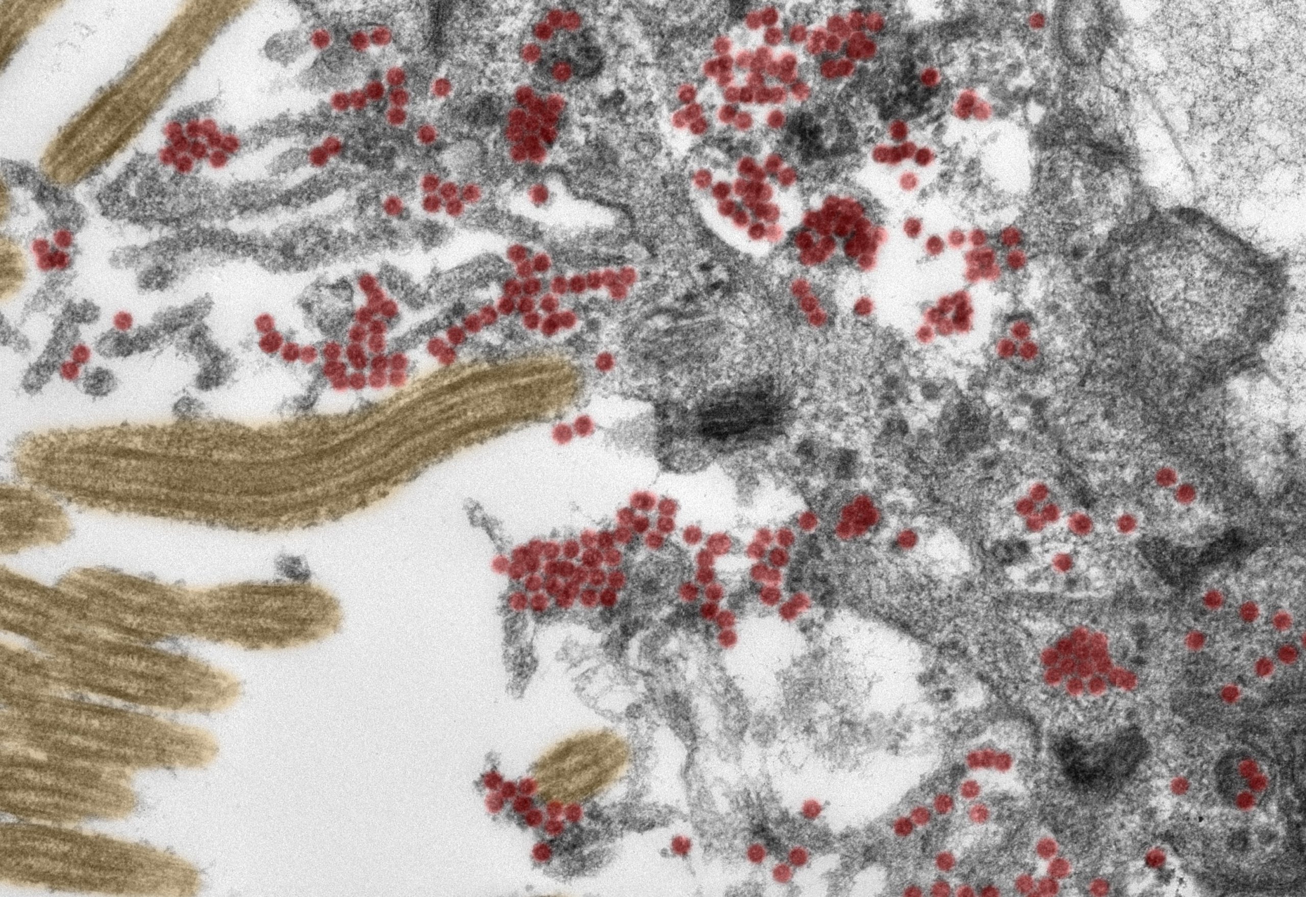
For the first time, researchers have been able to produce electron microscope images of intact coronavirus particles inside the olfactory mucosa.
It is now recognized that COVID-19 is not a purely respiratory disease.
In addition to affecting the lungs, SARS-CoV-2 can impact the cardiovascular system, the gastrointestinal tract, and the central nervous system.
The actions of COVID-19 on the brain: more than one in three patients exhibit loss or change in smell or taste, headaches, fatigue, dizziness, and nausea
More than one in three people with COVID-19 report neurological symptoms such as loss of, or change in, their sense of smell or taste, headaches, fatigue, dizziness, and nausea.
In some patients, the disease can even result in stroke or other serious conditions.
Until now, researchers had suspected that these manifestations must be caused by the virus entering and infecting specific cells in the brain.
But how does SARS-CoV-2 get there?
Under the joint leadership of Dr. Helena Radbruch of Charité’s Department of Neuropathology and the Department’s Director, Prof. Dr. Frank Heppner, a multidisciplinary team of researchers has now traced how the virus enters the central nervous system and subsequently invades the brain.
As part of this research, experts from the fields of neuropathology, pathology, forensic medicine, virology, and clinical care studied tissue samples from 33 patients (average age 72) who had died at either Charité or the University Medical Center Göttingen after contracting COVID-19.
Using the latest technology, the researchers analyzed samples taken from the deceased patients’ olfactory mucosa and four different brain regions.
Both the tissue samples and distinct cells were tested for SARS-CoV-2 genetic material and a ‘spike protein’ which is found on the surface of the virus.
The team provided evidence of the virus in different neuroanatomical structures that connect the eyes, mouth, and nose with the brain stem.
COVID-19 damage to the brain: the olfactory mucosa revealed the highest viral load
The olfactory mucosa revealed the highest viral load. Using special tissue stains, the researchers were able to produce the first-ever electron microscopy images of intact coronavirus particles within the olfactory mucosa.
These were found both inside nerve cells and in the processes extending from nearby supporting (epithelial) cells.
All samples used in this type of image-based analysis must be of the highest possible quality.

To guarantee this was the case, the researchers ensured that all clinical and pathological processes were closely aligned and supported by a sophisticated infrastructure.
“These data support the notion that SARS-CoV-2 can use the olfactory mucosa as a port of entry into the brain,” says Prof. Heppner.
This is also supported by the close anatomical proximity of mucosal cells, blood vessels, and nerve cells in the area.
“Once inside the olfactory mucosa, the virus appears to use neuroanatomical connections, such as the olfactory nerve, to reach the brain,” adds the neuropathologist.
“It is important to emphasize, however, that the COVID-19 patients involved in this study had what would be defined as severe disease, belonging to that small group of patients in whom the disease proves fatal.
It is not necessarily possible, therefore, to transfer the results of our study to cases with mild or moderate disease.”
How the virus moves on from the nerve cells remains to be fully elucidated.
“Our data suggest that the virus moves from nerve cell to nerve cell to reach the brain,” explains Dr. Radbruch.
She adds: “It is likely, however, that the virus is also transported via the blood vessels, as evidence of the virus was also found in the walls of blood vessels in the brain.”
SARS-CoV-2 is far from the only virus capable of reaching the brain via certain routes.
“Other examples include the herpes simplex virus and the rabies virus,” explains Dr. Radbruch.
Researchers also studied how the immune system responds to COVID-19 infection
In addition to finding evidence of activated immune cells in the brain and the olfactory mucosa, they detected the immune signatures of these cells in the cerebral fluid.
In some of the cases studied, the researchers also found tissue damage caused by stroke as a result of thromboembolism (i.e. the obstruction of a blood vessel by a blood clot).
“In our eyes, the presence of SARS-CoV-2 in nerve cells of the olfactory mucosa provides a good explanation for the neurologic symptoms found in COVID-19 patients, such as a loss of the sense of smell or taste,” explains Prof. Heppner.
“We also found SARS-CoV-2 in areas of the brain which control vital functions, such as breathing.
It cannot be ruled out that, in patients with severe COVID-19, the presence of the virus in these areas of the brain will have an exacerbating impact on respiratory function, adding to breathing problems due to SARS-CoV-2 infection of the lungs. Similar problems might arise about cardiovascular function.”
Article on COVID-19 infection of the brain published by the Charité – Universitätsmedizin Berlin
s41593-020-00758-5 (1)Read Also:
Can Proteins Predict How Ill A Patient Could Become With COVID-19?
Russia, MEDEVAC For Alexej Navalny Who Flies To The Charité Hospital In Germany