
Hollow foot: what it is and how to recognise it
Hollow foot is a structural alteration of the foot characterised by an increase in the longitudinal arch of the foot
It may depend on various causes and may sometimes represent the tip of the iceberg of more important neurological diseases that should not be underestimated.
For this reason it can be considered as a pathology of multidisciplinary competence because it involves various specialists including the orthopaedist, radiologist, neurologist and geneticist.
Types of hollow foot
The hollow foot can be classified from a morphological point of view in 3 situations
- posterior hollow foot, i.e. affecting the back of the foot. It is mainly due to verticalisation of the calcaneus;
- Anterior hollow foot, i.e. involving the front part of the foot. It is mainly due to plantarflexion of the forefoot;
- Mixed hollow foot, i.e. affecting both parts of the foot.
The causes of hollow foot
This deformity has an aetiological classification, which can be traced back to 3 particular causes:
- idiopathic-congenital hollow foot;
- hollow foot due to neuromuscular causes;
- hollow foot due to trauma or injury.
Idiopathic-congenital hollow foot
The idiopathic-congenital hollow foot, also defined as physiological-familial, is due to causes attributable to members of one’s own household who also have a hollow foot.
It is a pathology that usually affects both feet (symmetrical), is not developmental and appears from childhood.
Sometimes this type of hollow foot also corrects itself as the child grows up, over the years, because it does not develop in a worsening direction, as happens with the other types.
Neuromuscular hollow foot
The neuromuscular hollow foot is the most serious in a certain sense because it is the clinical evidence of a latent neurological pathology such as, for example, a hereditary sensory-motor neuropathy or Charcot-Marie-Tooth disease (CMT), expressions of hereditary neurological pathologies which absolutely must be identified and treated as soon as possible.
In addition to these, there may also be diseases of the central nervous system such as meningocele or myelo-meningocele, or ‘acquired’ diseases of the nervous system such as poliomyelitis, infantile cerebral palsy or sciatic nerve lesions.
Hollow foot due to trauma or injury
The third type of hollow foot is caused, for example, by complex fractures, which may mainly affect the midfoot (i.e. the central part of the foot) or the metatarsals, or also by fractures of the leg.
In addition to these there are also tendon lesions, such as for example the tibialis anterior or peroneus brevis, because a muscular imbalance is generated which highlights the deformity in the hollow foot; or burns, especially if they occur in infancy, in the paediatric age, which may lead to retraction of the soft skin tissues and give evidence of a hollow foot.
The so-called compartmental syndromes may also be included among the causes of hollow foot in the third type.
How does hollow foot manifest itself
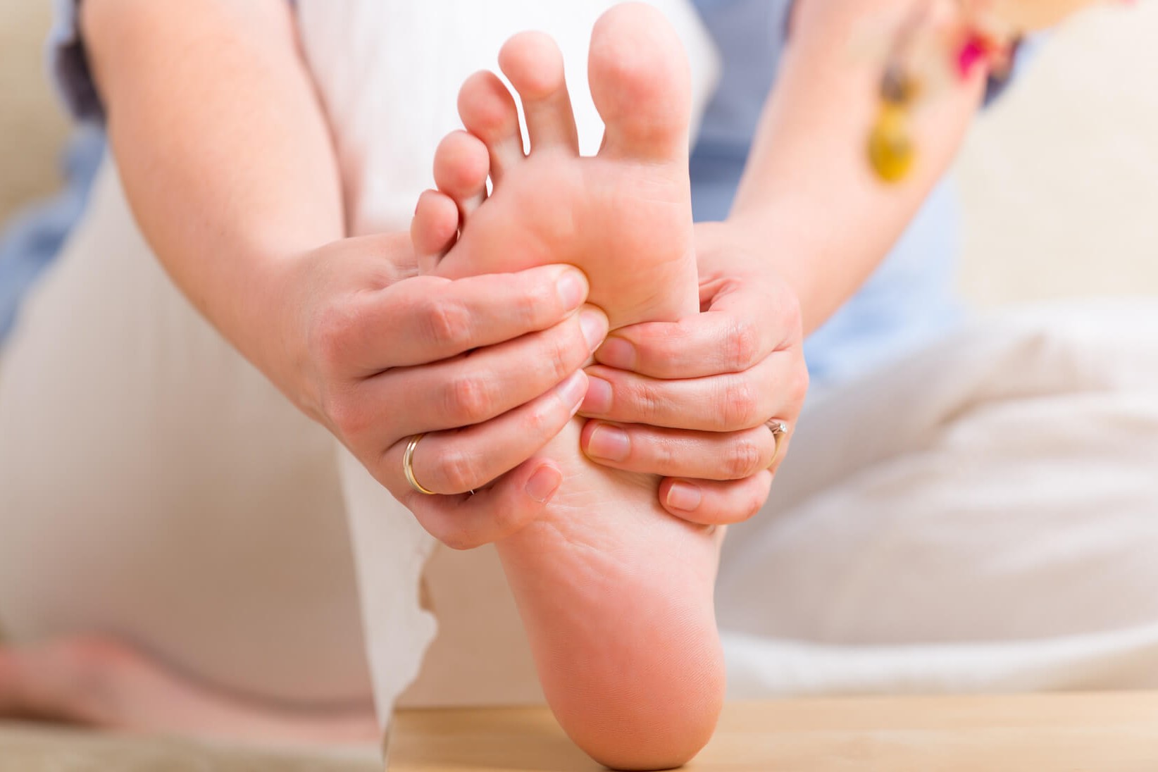
A hollow foot can be recognised by the shape of the foot, which has an increased plantar arch.
If there are already similar cases of hollow foot in the family, without necessarily leading to neuromuscular pathologies or secondary pathologies, the child inherits this disorder from the mother’s or father’s side.
Often, simple corrective measures such as orthotics are useful.
A person usually goes to a specialist because he or she presents various symptoms such as, for example, metatarsalgia, i.e. pain in the sole of the foot, perhaps with the appearance of hyperkeratosis (callosity) in the anterior plantar area, or ankle instability, one of the possible causes of which is the hollow foot, or, in cases of neuromuscular hollow foot, weakness and fatigue.
The appearance of claw toes may also be a warning sign.
Diagnosis of hollow foot
When visiting the doctor, the patient is examined either lying on the couch or standing; he is made to walk and an analysis of gait is carried out which provides a broader view of the functioning of hollow feet, i.e. with support occurring mainly on the outer edge (in supination).
If necessary, one can proceed with baropodometry, which consists of having the person walk on a platform, detecting any maladjustment of the foot, or situations in which the activated muscles work or do not work.
It is also possible to note areas of the foot that are leaning too much, such as at the front or at the back, the so-called tripod effect.
In support of the clinic and a very accurate anamnesis, instrumental examinations can be carried out to further confirm the diagnosis.

These include
- X-ray of the feet in load, more frequent;
- CT scan of the weight-bearing feet, where required;
- magnetic resonance imaging, rarer;
- ultrasound, rarer.
Diagnosis of hollow foot with neuromuscular cause
Obviously not all hollow feet need to undergo CT scans of this type.
In the case of hollow feet with mainly neuromuscular causes, it is important to carry out an electromyography.
In cases of neuromuscular pathologies such as muscular dystrophies, Charcot-Marie-Tooth and others, a DNA examination can be carried out or even a blood test to evaluate the so-called CPK (creatine phosphokinase) enzymes, which is useful for investigating possible myopathies or, in even more severe cases, a muscle biopsy.
A neurological evaluation is also essential in these circumstances.
How is the disease treated?
Treatment can be
- conservative;
- surgical.
Conservative treatment with exercises
From the conservative point of view, which is always the first step unless it is a neuromuscular hollow foot, we can proceed with physiokinesitherapy.
When the hollow foot is mild, and therefore does not present a progression towards deformity, gymnastics can be used with the help of a physiotherapist (following a physiatric assessment).
This practice is useful for several aspects
- prevention of contractures
- preservation of proprioception, especially in adolescence;
- strengthening the capsular ligament structures of the ankle in the event of instability.
Insoles
As already mentioned, another conservative intervention is the podiatric approach through insoles and digital silicone orthoses (orthotics), which are basically useful to avoid overloading and prevent calluses from forming.
They act by distributing the pressure of the support, increasing the surface area and avoiding possible conflicts with the footwear.
The footwear must be adapted to the size of the foot, continues the orthopaedist.
Surgical treatment
The treatment is surgical and intervenes on the bone in various ways:
- osteotomy of the heel or metatarsals, an operation which fractures the bone in order to realign it;
- arthrodesis, an operation to block one or two joints, with the involvement of the big toe or claw toes. The big toe can also sometimes go into a jaw (flexed toe).
In addition to operations on the bones, operations can be carried out on soft parts such as tendons through tenodesis or tendon transpositions.
The important thing is to try to recover a certain muscular balance.
Read Also:
Emergency Live Even More…Live: Download The New Free App Of Your Newspaper For IOS And Android
Orthopaedics: What Is Hammer Toe?
(Also) Occupational Diseases: All Causes And Remedies Of Plantar Fasciitis
Pain In The Sole Of The Foot: It Could Be Metatarsalgia