
PET: what it is for and how to take the exam
PET (Positron Emission Tomography) is a diagnostic method in Nuclear Medicine that, in response to a precise clinical question, can provide extremely precise information on pathologies of organs or tissues of the body
PET is used in particular in Oncology, both in the diagnostic phase to identify diseased tissue and staging a tumour, and during follow-up after surgery or radiotherapy to assess the progress of a treatment.
Also useful in other areas, in Neurology PET is used to differentiate Alzheimer’s from other dementias, in Cardiology it analyses the flow of the heart and the vitality of its tissues, and in Orthopaedics it is used to assess the status of vertebral infections and infected prostheses.
How PET works
PET is a non-invasive examination that poses no risk to the patient.
It is carried out using a radiopharmaceutical, administered intravenously into the forearm, and consisting of a molecule (or tracer) that maps the pathological process of interest and an atom that emits positrons with a short half-life.
Like a light bulb, the drug ‘illuminates’ the cells to which it binds, highlighting the presence of any pathologies and allowing their complete and precise staging.
Each of the drugs used in the PET scan consists of specific molecules that are only recognised by the tissues to be analysed, thus ensuring a high degree of accuracy.
Following the administration of the drug, the patient must wait for a period of time, which can vary from a few minutes to an hour, before undergoing the examination, to allow the radiopharmaceutical to distribute itself adequately in the body and reach the target tissue.
At the end of this wait, the actual examination can begin. It will last about 30 minutes, with small variations possible, depending on the tissue to be investigated.
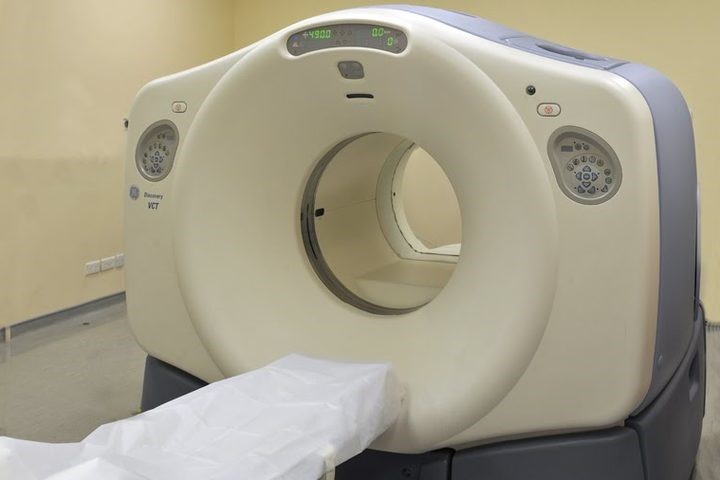
The patient is asked to lie supine and motionless in an open ring-shaped machine, the tomograph.
This instrument, capable of recording the radiation emitted by the drug, will examine the body by reproducing the recorded images on a computer, which will then be evaluated by the specialist doctor.
Immediately before the PET images are taken, a CT scan is always performed with the same tomograph, which is necessary for correct reconstruction of the images and anatomical localisation of any changes visible on the PET scan.

In selected cases, a CT scan with contrast medium is performed, so that two examinations are obtained in one diagnostic session.
PET examination preparation
To prepare for the PET scan, it is sufficient to fast for 6 hours. In the case of FDG PET the night before, it is recommended to maintain a carbohydrate-free diet.
The patient is almost always asked to empty his bladder before the test in order to allow proper visualisation of the organs.
It is not necessary to undress for the PET scan, but comfortable clothing should be worn.
In addition, the patient should not have any metal objects on him or her, as these may interfere with the correct performance of the test.
Once the test has been completed, the patient can leave the ward without having to take any special precautions.
The radiation emitted by the radiopharmaceuticals used has a very short half-life and is almost completely eliminated during the stay in the ward.
The examination is, however, contraindicated for pregnant women in order to avoid unnecessary radiation to the foetus, bearing in mind that a CT scan is also always performed.
Read Also:
Psychosis Is Not Psychopathy: Differences In Symptoms, Diagnosis And Treatment