
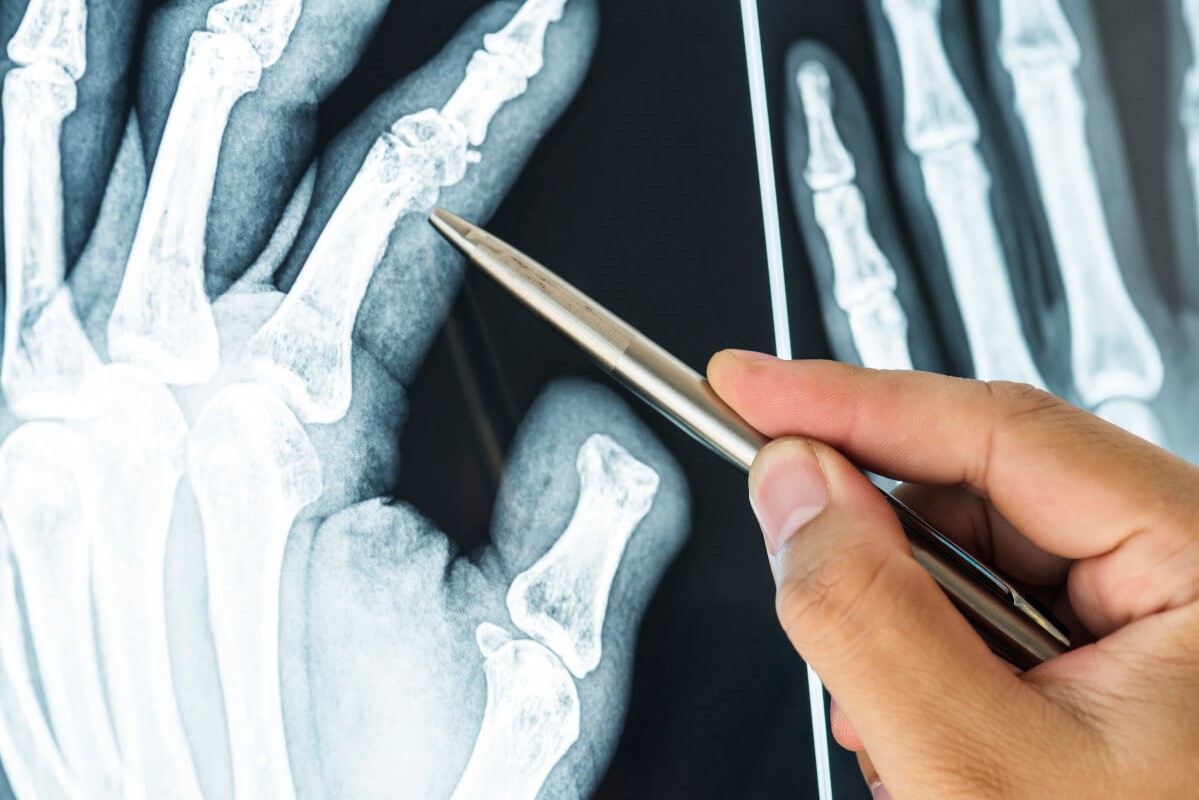
What is hand radiography (hand X-ray)?
Radiography (or X-ray) of the hand: the X-ray examination of the hand is a radiological examination carried out using X-rays, which are able to pass through the human body and imprint themselves photographically on a film
The result is an image that shows bones and skeleton differently from soft tissues (muscles and skin), because bones are more compact structures and appear lighter than other tissues.
Currently, X-ray techniques produce a digital image that is either scanned (computed radiography) or processed through a detector (digital radiography)
Although we still refer to the X-ray ‘plate’ in common usage, today the examination results are delivered to the patient in digital format, usually on CD and DVD.
What is hand X-ray used for?
The examination, which is quick and safe, is performed in cases of:
- Fracture, both in cases of trauma fracture and fractures caused by bone weakness due to existing injuries.
- To evaluate the healing of the bone after a fracture (to show the formation of bone callus) or after surgical interventions involving the bone (prostheses, synthesis means, nails, etc.).
- To exclude or check for tumour diseases (benign or malignant focal lesions).
- In preparation for surgery (e.g. for a prosthetic implant) and to monitor its effects over time.
- In the case of degenerative processes that lead to changes in bones and joints, such as arthrosis, causing chronic pain or movement problems.
- In the case of inflammatory processes, such as arthritis, or infectious processes.
- To reveal the state of wear and tear of joint cartilage.
- In the case of tendon problems (tendinitis) and ligament problems, even if the X-ray cannot obtain images, it can identify indirect signs of a tendon injury such as calcifications.
- In children, x-rays of the hand, together with x-rays of the wrist, can be used in growth defects to conduct the so-called bone age study and to know the degree of skeletal maturation.
- Are there any rules of preparation for hand X-ray?
- There are no special preparation rules.
There is no fasting, no water intake and no need to suspend ongoing drug therapies.
Who can take an X-ray of the hand?
The examination can be performed by anyone, except pregnant women.
Is hand X-ray painful or dangerous?
The X-ray examination is easy to perform, non-invasive and safe.
There is no risk of allergic reactions because no contrast medium is used.

The effects of radiation on the body are well known, as are the potential risks, but the radiation doses used today and the exposure time have been significantly reduced compared to previous years, which is beneficial for patients.
The use of the latest generation of digital X-ray equipment and new shielding procedures to protect the most radiosensitive parts of the body have further reduced the risks.
How does hand X-ray work?
The examination takes about 5 minutes.
Longer times are only required for particularly complex examinations or when more images are needed.
The examination is conducted by placing the area under examination and the patient at the X-ray machine in a protected room.
The patient can go home immediately after the examination and does not need to be accompanied.
Read Also:
Intraosseous Access, A Life-Saving Technique In Emergency Shock Management
Electromyography (EMG), What It Assesses And When It Is Done
Dislocation Of The Shoulder: How To Reduce It? An Overview Of The Main Techniques