What is an ECG and when to do an electrocardiogram
The electrocardiogram is an examination that makes it possible to diagnose numerous heart diseases. The expert explains how it works and what it consists of
According to data from the Ministry of Health, cardiovascular disease is still the leading cause of death in Italy, accounting for 34.8% of all deaths.
Many cardiovascular diseases can be diagnosed with basic, first-level tests, including the electrocardiogram.
What is the Electrocardiogram (ECG)?
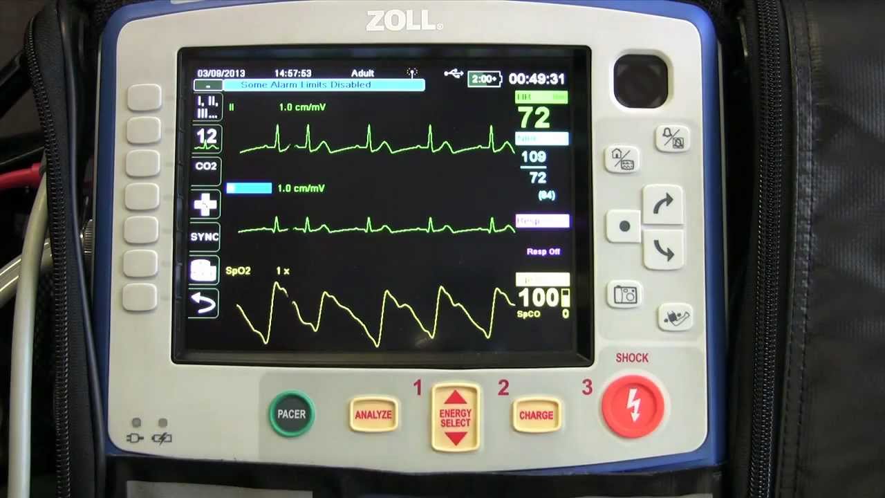
The electrocardiogram (ECG) is an examination that records the intrinsic electrical activity of myocardial fibres.
In simple terms, it is a practical, easily repeatable and inexpensive method of recording the electrical activity of the heart to observe whether mechanical or bioelectrical disorders are present.
What is the purpose of the electrocardiogram (ECG)?
The electrocardiogram allows the cardiologist to diagnose a number of cardiac disorders and pathologies including:
- arrhythmias: changes in heart rhythm: the heart beats irregularly, too slowly or too fast. The diagnosis of arrhythmias is very important, as they are often asymptomatic and can lead to cardiac arrest and sudden death;
- ischaemia and/or infarction: the ECG can detect cardiac distress caused by a reduction in the flow of blood to the heart (ischaemia) caused by a narrowing of a coronary artery, which can lead to myocardial infarction (death of heart tissue);
- congenital or acquired alterations and physical disorders of the heart cavities such as valvulopathies, ventricular hypertrophy, dilated cardiomyopathies, etc;
- electrolyte disorders: excess or deficient concentration of blood electrolytes, leading to an alteration in cardiac rhythm;
- toxic effects of certain drugs: which can cause damage to the heart muscle.
The ECG also allows the evaluation of the functioning of pacemakers and other internal devices such as implantable defibrillators.
ECG EQUIPMENT? VISIT THE ZOLL BOOTH AT EMERGENCY EXPO
Symptoms of heart disease to watch out for
Assuming that some heart diseases can be asymptomatic before very serious events such as cardiac arrest, the symptoms to look out for and which may indicate heart disease are very variable, but can consist of:
- absence of pulse;
- chest pain
- easy fatigability;
- sense of weakness (asthenia);
- frequent swelling of the lower limbs;
- prolonged headaches and dizziness;
- shortness of breath (dyspnoea);
- palpitation;
- feeling of an irregular heartbeat;
- frequent fainting (lipothymia).
When to perform an electrocardiogram
The electrocardiogram is a very simple diagnostic test to carry out, which is indicated in cases where:
- the symptoms mentioned above are present, which may be due to heart disease;
- there are family risk factors, which are very important when assessing the patient’s state of health, as various heart diseases may have a family predisposition;
- there is a need to complete the clinical-cardiocirculatory picture of a patient who, for example, is to undergo surgery;
- it is necessary to obtain certification for sporting activity, including competitive sport, in the context of clinical assessments to ascertain the state of health of the athlete;
- you need to assess the development of heart disease over time or check the effectiveness of treatment.
How the examination is carried out
The ECG lasts a few minutes.
Ten electrodes are placed on the patient’s body (arms, legs and chest) to record the heart’s electrical activity.
The electrocardiograph then reproduces this in a trace which is evaluated by the specialist.
There is no electrical stimulation and there are no particular contraindications to the examination, which is painless and non-invasive.
How often should an ECG be performed?
It is up to the specialist to decide how often to carry out medical check-ups and an electrocardiogram, depending on the results of the examination and the presence or absence of pathologies or risk factors.
From the age of 40 onwards, it would be advisable to have them every two years and after 50 at least once a year.
Types of electrocardiogram
Depending on the symptoms and the type of problem highlighted or suspected, there are also other types of ECG that can be performed:
- Basal ECG (at rest): this is the classic examination method, with the patient lying supine on a couch and the electrodes placed on his body;
- Holter dynamic ECG: it is carried out with a small portable electrocardiograph that allows the recording of cardiac activity continuously for 24 hours, highlighting phenomena (arrhythmias, coronary insufficiency, etc.) that would otherwise be unknown;
- Exercise ECG: is the evaluation of the heart under physical stress with real-time monitoring of the electrocardiogram and blood pressure. It makes it possible to observe the behaviour of blood pressure and to highlight the onset of arrhythmias and myocardial ischaemia phenomena during physical work;
- Loop recorder: this is carried out by subcutaneous application of a device that records cardiac electrical activity during the day and transmits the information to the operations centre at night. This investigation can last for several months and is indicated to assess the existence of rare but potentially serious or dangerous phenomena such as malignant arrhythmias, syncopes, etc.
- Other cardiological examinations
The electrocardiogram is one of the basic and fundamental cardiac examinations, but it is not the only one that allows assessment of cardiac function.
In addition to this, we should also mention
- colordoppler echocardiogram: a sophisticated ultrasound scan of the heart, carried out with an ultrasound probe in the event that heart damage or defects are suspected;
- resting and exercise myocardial scintigraphy: depending on the type of examination indicated, after an exercise test or a pharmacological provocative test, a weakly radioactive drug is injected into the patient. The images acquired by a piece of equipment, called a gamma camera, provide information on how the blood flows into the myocardium (the muscle area) at rest or under stress, to allow assessments of cardiac function;
- coronary angiography (virtual coronary angiography, coronaro tc): this is a computerised axial tomography (CT) scan with contrast medium, which can produce high-definition 3D images of the coronary arteries and thus non-invasively assess the presence of any narrowing (stenosis);
- coronarography: this is an examination involving the administration of a contrast medium used to make the coronary arteries visible on x-rays, so as to assess the presence of any stenosis;
- myocardial resonance imaging (MRI): this test uses magnetic resonance imaging to produce images that assess the anatomical structures of the heart, particularly the myocardium.
Read Also:
ST-Elevation Myocardial Infarction: What Is A STEMI?
ECG First Principles From Handwritten Tutorial Video
ECG Criteria, 3 Simple Rules From Ken Grauer – ECG Recognize VT


