
Diagnostic and operative hysteroscopy: when is it necessary?
Hysteroscopy is an outpatient endoscopic test that does not require analgo-anesthesia and allows us to investigate inside the uterine cavity using an instrument called a hysteroscope
This is a thin, rigid tube a few millimetres in diameter, equipped with optical fibres through which light travels, which is introduced inside the uterus, through the vagina.
When to undergo hysteroscopy?
Diagnostic hysteroscopy is indicated especially in the presence of abnormal uterine bleeding in the fertile period, in pre- and post-menopause, in patients undergoing post-neoplasm breast drug therapy or undergoing replacement treatment for menopause.
It is important to undergo diagnostic hysteroscopy in cases of couple infertility (which should always be performed prior to any access to AMP techniques), in suspected uterine malformations, for post-surgery hysteroscopic uterine cavity checks, and in cases of post-abortion or post-partum chorioplacental remnants.
Operative hysteroscopy, on the other hand, is used in the presence of
- intrauterine adhesions
- uterine malformations such as uterine septum
- endometrial polyps
- submucosal uterine fibroids
- intrauterine foreign bodies, such as an IUD whose filament has ascended into the uterine cavity.
Stages of hysteroscopy: what to do before the test
Diagnostic hysteroscopy requires no special preparation. In the case of operative hysteroscopy, preparation requires fasting from midnight on the day before surgery.
On the day of the operation, antibiotic prophylaxis is carried out and the patient is asked to empty her bladder.
Anaesthesia is necessary. In this case, the patient must have undergone the following investigations: blood tests, electrocardiogram and chest X-ray (if age > 50 years).
Stages of hysteroscopy: what to do during and after
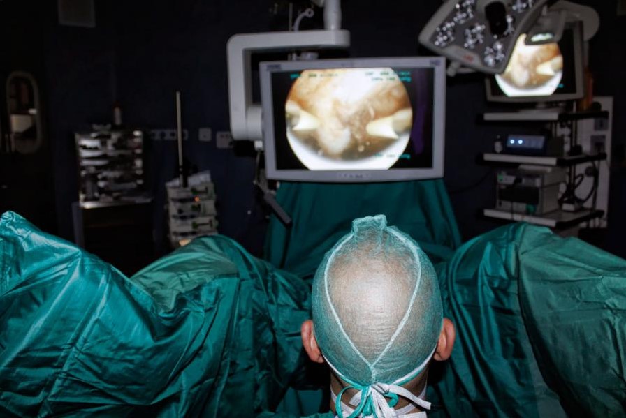
Access to the uterine cavity is by vaginoscopic, atraumatic technique: the hysteroscope is introduced, through the vagina, into the cervical canal until it reaches the uterine cavity, which is distended with a gaseous or liquid medium in order to allow it to be seen.
In the case of surgical hysteroscopy, miniaturised instruments, such as scissors or forceps, can be introduced through the hysteroscope, or the resectoscope is used to cut and coagulate through a source of electrical energy.
The diagnostic hysteroscopy lasts only a few minutes; at the end of the procedure, the hysteroscope is removed and the distention medium flows out of the uterine cavity, which returns to its original size.
No sutures or dressings are required.
The diagnostic hysteroscopy causes no particular discomfort and the patient quickly resumes her activities.
In some cases, she may experience cramp-like pain similar to menstruation and a modest dripping of blood (spotting), both of which quickly disappear.
Treatment following hysteroscopy varies from patient to patient.
In any case, a gynaecological examination is required after one month to assess the advisability of pharmacological treatment or further surgery.
Hysteroscopy: contraindications and risks
Contraindications to performing hysteroscopy are:
- the presence of an ongoing pregnancy
- the presence of an ongoing or recent pelvic infection
- carcinoma of the cervix.
Diagnostic hysteroscopy is almost risk-free and complications are very rare.
Operative hysteroscopy, on the other hand, carries the following risks
- nausea and vomiting as a result of anaesthesia
- perforation of the uterus possible, but infrequent;
- injury to abdominal organs very rare;
- cardiac arrest and/or pulmonary oedema, very rare occurrences;
- cardiovascular overload during the operation may be a complication related to the fluid used to distend the uterine cavity, an event that can be serious, but rare and well-predictable.
Read Also
Emergency Live Even More…Live: Download The New Free App Of Your Newspaper For IOS And Android

Techniques And Instruments For Performing Hysteroscopy
The Use Of Outpatient Hysteroscopy For Early Diagnosis
Utero-Vaginal Prolapse: What Is The Indicated Treatment?
Pelvic Floor Dysfunction: What It Is And How To Treat It
Pelvic Floor Dysfunction: Risk Factors
Salpingitis: Causes And Complications Of This Fallopian Tube Inflammation
Hysterosalpingography: Preparation And Usefulness Of The Examination
Gynaecological Cancers: What To Know To Prevent Them
Total And Operative Hysterectomy: What They Are, What They Involve
Vulvodynia: What Are The Symptoms And How To Treat It
What Is Vulvodynia? Symptoms, Diagnosis And Treatment: Talk To The Expert
Accumulation Of Fluid In The Peritoneal Cavity: Possible Causes And Symptoms Of Ascites
Accumulation Of Fluid In The Peritoneal Cavity: Possible Causes And Symptoms Of Ascites
What’s Causing Your Abdominal Pain And How To Treat It
Pelvic Varicocele: What It Is And How To Recognise The Symptoms
Can Endometriosis Cause Infertility?
Transvaginal Ultrasound: How It Works And Why It Is Important
Candida Albicans And Other Forms Of Vaginitis: Symptoms, Causes And Treatment
What Is Vulvovaginitis? Symptoms, Diagnosis And Treatment
Vaginal Infections: What Are The Symptoms?
Chlamydia: What Are The Symptoms And How To Treat It
Chlamydia, Symptoms And Prevention Of A Silent And Dangerous Infection
Miscarriage: Causes, Diagnosis And Treatment
Diagnostic And Operative Hysteroscopy: Preparation And Importance Of Gynaecological Examinations
Urethrocistoscopy: What It Is And How Transurethral Cystoscopy Is Performed
Uterine Fibroid Embolization: What It Is And How To Treat It