
Gastroscopy: what the examination is for and how it is performed
Gastroscopy, or examination of the stomach, is useful for confirming or ruling out diseases such as gastritis, ulcers or tumours of the oesophagus, stomach or duodenum
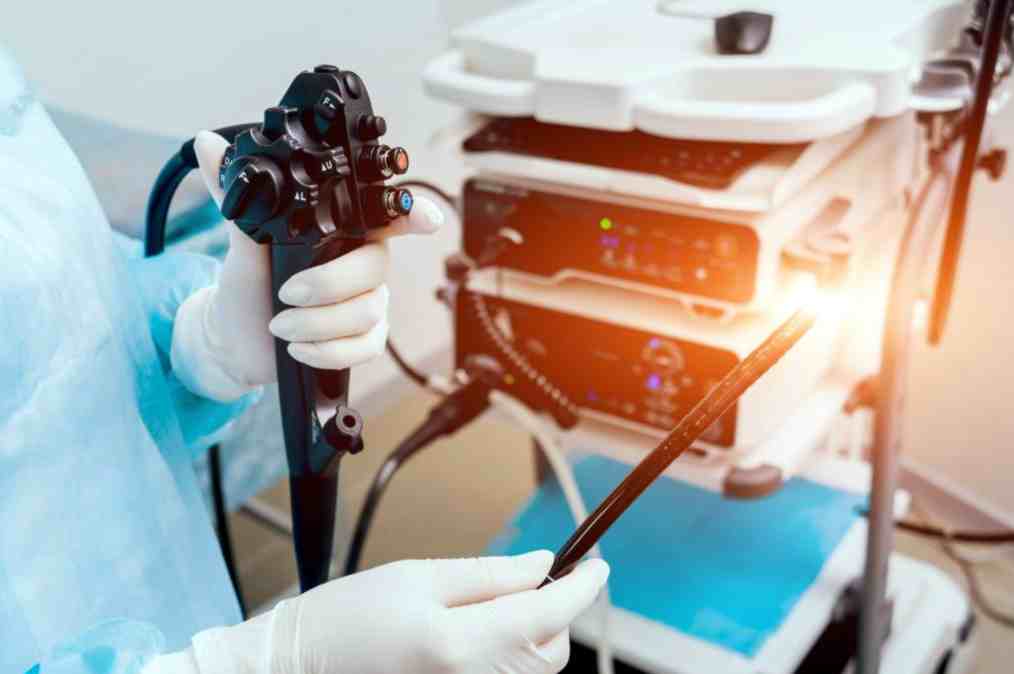
The examination uses an instrument called a gastroscope that ‘looks’ inside the oesophagus, stomach and duodenum, the first part of the intestine.
The gastroscope is a tube about 1 cm in diameter, with a small light and a camera at the end, which is controlled by the operator using knobs.
The image from the camera is then sent to the screen. The endoscope has channels called operators, through which instruments (biopsy forceps) can be inserted to obtain tissue samples for analysis.
When a gastroscopy is needed
A gastroscopy can be performed when you have one or more symptoms that can be traced back to gastrointestinal diseases such as:
- chronic or recurrent heartburn;
- prolonged nausea or vomiting
- stomach pain;
- difficulty swallowing
- black or bloody stools;
- weight loss for no apparent reason
- suspected peptic ulcer;
- suspected cancer of the oesophagus or stomach;
- suspected malabsorption – as in the case of coeliac disease;
- a check-up after stomach surgery.
Gastroscopy: how to prepare?
Before the gastroscopy, the specialist will explain the procedure and its risks to the patient, including all the information about the medication that will be given to make the examination more comfortable.
The patient should inform the doctor of any medication he or she is taking regularly, so that the doctor can decide whether to discontinue it before the procedure.
It may be necessary to stop taking certain medications, such as anticoagulants, a few days before the gastroscopy.
Your doctor will also tell you how many hours before the gastroscopy you should stop eating and drinking.
If the gastroscopy requires sedation, patients will not be allowed to drive or operate machinery for 12-24 hours after the procedure – the exact time will depend on the medication used.
It would be preferable for the patient to go home accompanied by someone after the gastroscopy.
How a gastroscopy takes place
A gastroscopy usually takes a few minutes.
The patient is asked to lie on his left side and is asked to hold a small mouthpiece between his teeth so that he can keep his mouth open and prevent him from biting the gastroscope.

Before starting the examination, an anaesthetic spray can be used to numb the throat.
The patient is then asked to swallow so that the gastroscope can enter the oesophagus, after which the instrument is slowly pushed into the stomach and then into the first part of the duodenum.
During the examination, air will also be inserted to distend the viscera to be examined and water may be used to clean the walls.
Both air and water may be removed during the procedure.
What is gastroscopy used for
Using video images, the specialist examines the lining of the oesophagus, stomach and duodenum to look for redness and possible signs of inflammation or lesions such as ulcers or tumours.
Through this examination, possible sources of bleeding can also be identified and haemostasis performed to stop the bleeding.
If necessary, tissue samples will be collected during the procedure and analysed under a microscope.
At the end of the gastroscopy, the patient will receive a written report and possibly photographic documentation.
What are the risks of gastroscopy?
The examination may result in swelling in the abdomen, a sore throat or numbness in the mouth caused by the anaesthetic spray.
Only in rare cases do the sedatives used lead to complications such as breathing problems or cardiovascular problems.
Read Also:
Emergency Live Even More…Live: Download The New Free App Of Your Newspaper For IOS And Android
Gastroesophageal Reflux: Causes, Symptoms, Tests For Diagnosis And Treatment
Esophageal Achalasia, The Treatment Is Endoscopic