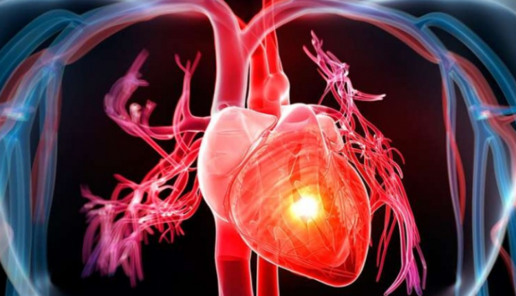
Heart failure: causes, symptoms, tests for diagnosis and treatment
Heart failure is one of the most common cardiopathies in the over-65s. It is characterised by the inability of the heart to perform its pump function, resulting in insufficient blood supply to the rest of the body and “stagnation” of blood upstream of the dysfunctional heart chambers, which leads to “congestion” of the affected organs. This is also referred to as heart failure
What is heart failure? What does it consist of?
Heart failure is a chronic condition whose frequency in Italy is about 2%, but it becomes progressively more frequent with age and in the female sex, reaching 15% in both sexes in the over 85s.
Due to the general ageing of the population, it is currently the cardiovascular disease with the highest incidence (1-5 new cases per 1000 subjects/year) and prevalence (over 100 cases per 1000 subjects over 65 years) and the main cause of hospitalisation in people over 65 years of age.
Systolic decompensation and diastolic decompensation
The heart receives venous blood from the periphery (via the right atrium and ventricle), promotes oxygenation by introducing it into the pulmonary circulation, and then, via the left atrium and ventricle, pushes the oxygenated blood into the aorta and then into the arteries for transport to all the organs and tissues of the body.
An initial distinction can therefore be made between:
- Systolic decompensation, in the presence of a reduced capacity of the left ventricle to excrete blood;
- Diastolic decompensation, in the presence of impaired left ventricular filling.
Since left ventricular function is commonly assessed by the so-called ejection fraction (percentage of blood pumped into the aorta at each contraction (systole) of the left ventricle), usually calculated by echocardiogram, a more precise distinction between:
- Preserved ejection fraction (or diastolic) decompensation, in which the ejection fraction is greater than 50%.
- Reduced ejection fraction (or systolic) decompensation, in which the ejection fraction is less than 40%.
- Slightly reduced ejection fraction decompensation, where the ejection fraction is between 40 and 49%.
This classification is important for the development of increasingly targeted therapies (as we shall see, there are currently only proven therapies for reduced ejection fraction decompensation).
Heart failure: What are the causes?
The cause of heart failure is usually damage to the myocardium, the heart muscle, which can be caused, for example, by a heart attack or by excessive stress caused by uncontrolled hypertension or valve dysfunction.
The electrocardiogram of many decompensated patients may show a left bundle branch block (BBS), an alteration in the propagation of the electrical impulse that can change the mechanics of the heart, causing a dyssynchrony of contraction and, consequently, worsening cardiac contractile activity.
Heart failure: risk factors
In more detail, the following are risk factors for decompensation with reduced ejection fraction
- ischaemic heart disease (in particular previous myocardial infarction)
- valvular heart disease
- hypertension.
On the other hand, risk factors for decompensation with preserved ejection fraction are
- diabetes
- metabolic syndrome
- obesity
- atrial fibrillation
- hypertension
- female sex.
What are the symptoms of heart failure?
In the early stages of heart failure, symptoms may be absent or mild (such as breathlessness after strenuous exercise).
Heart failure, however, is a progressive condition, whereby symptoms gradually become more noticeable, leading to the need to seek medical attention or sometimes necessitating hospitalisation.
Symptoms, a consequence of reduced blood supply to organs and tissues and ‘stagnation’ of blood upstream of dysfunctional cardiac chambers with ‘congestion’ of the affected organs, may include:
- Dyspnoea, i.e. shortness of breath, caused by the accumulation of fluid in the lungs: initially it appears after intense exertion, but gradually also after mild exertion, at rest and even lying supine during sleep (decubitus dyspnoea), interrupting night-time rest and forcing one to sit up.
- Edema (swelling) in the lower limbs (feet, ankles, legs), also caused by a build-up of fluid.
- Abdominal swelling and/or pain, again caused by fluid accumulation, in this case in the viscera.
- Asthenia (tiredness), caused by reduced blood supply to the muscles.
- Dry cough, due to accumulation of fluid in the lungs.
- Loss of appetite.
- Difficulty concentrating, caused by reduced blood supply to the brain, and, in severe cases, confusion.
Heart failure: levels of severity
Based on the symptoms that physical activity generates and, therefore, the degree to which it is restricted, the New York Heart Association has defined four classes of increasing severity (from I to IV) of heart failure:
- Asymptomatic patient: habitual physical activity does not cause fatigue or dyspnoea.
- Mild heart failure: After moderate physical activity (e.g. climbing a couple of flights of stairs or just a few steps with a weight), dyspnoea and fatigue are experienced.
- Moderate to severe heart failure: dyspnoea and fatigue occur even after minimal physical activity, such as walking less than 100 m on level ground at a normal pace or climbing a flight of stairs.
- Severe heart failure: asthenia, breathlessness and fatigue occur even at rest, sitting or lying down.
Diagnosis: a cardiological examination
Obtaining an early diagnosis of heart failure is important to better manage this chronic condition, slowing down its progression and thus helping to improve the patient’s quality of life.
However, diagnosing heart failure is not always easy: the symptoms often fluctuate, varying in intensity as the days go by.
Moreover, as we have seen, these are non-specific symptoms, which patients, especially elderly patients and those already struggling with other illnesses, tend to underestimate or attribute to other causes.
On the other hand, the presence of dyspnoea and/or oedema in individuals with risk factors for heart failure should prompt a specialist cardiological examination.
What tests should be done to diagnose heart failure?
The diagnostic examination for heart failure includes a history (i.e. gathering information about the patient’s medical history and symptoms) and a preliminary physical examination. The specialist may then ask for some additional investigations (laboratory and instrumental tests), including
- electrocardiogram
- echocardiogram
- magnetic resonance imaging of the heart with contrast medium
- blood dosage of natriuretic peptides (molecules produced mainly by the left ventricle; normal blood levels generally rule out decompensation).
More invasive tests, such as cardiac catheterisation and coronarography, may also be required.
How is heart failure treated?
Heart failure is a chronic condition that requires a multidisciplinary approach in order to reduce symptoms, slow down the progression of the disease, reduce hospital admissions, increase patient survival and improve quality of life.
In addition to early diagnosis, the active role of the patient and the collaboration between the multidisciplinary team and the family doctor are valuable.
The main treatment options include:
- Lifestyle changes, which include:
- Reducing salt consumption;
- Regular aerobic physical activity of moderate intensity (e.g. 30 minutes of walking at least 5 days a week);
- Limiting fluid intake;
- Self-monitoring, i.e. daily monitoring of body weight, blood pressure, heart rate, possible presence of oedema.
- Pharmacological therapy, with several drugs in combination including:
- Drugs blocking the renin-angiotensin-aldosterone system (ACE inhibitors, sartans and antialdosteronic drugs);
- Drugs that antagonise the sympathetic nervous system (beta-blockers, such as carvedilol, bisoprolol, nebivolol and metoprolol);
- Neprilysin inhibitor drugs (such as sacubitril);
- Sodium-glucose cotransporter inhibitors.
- Cardiac resynchronisation therapy (in combination with medication, if there is a disorder of electrical impulse conduction, such as left bundle-branch block): requires the implantation of electrical devices (pacemakers or biventricular defibrillators), to resynchronise cardiac contraction. Together with medication, the devices can slow down the progression of the disease and sometimes lead to normalisation of the left ventricular ejection fraction.
- Surgical interventions (such as surgical or percutaneous correction of valve disease, surgical or percutaneous myocardial revascularisation, up to implantation of ‘artificial hearts’ and heart transplantation).
It should be pointed out that the above-mentioned drugs and resynchronisation therapy have only proved effective in systolic decompensation or reduced ejection fraction. In particular, the first two categories of drugs mentioned above, i.e. renin-angiotensin-aldosterone system blockers (ACE inhibitors, sartans and anti-aldosteronic drugs) and those that antagonise the sympathetic nervous system (beta-blockers), are still the first-line therapy for this condition.
These have been shown to change the history of the disease, reducing mortality and morbidity by acting on the negative interactions between hyper-activation of the sympathetic nervous system and the renin-angiotensin-aldosterone system and the progression of ventricular dysfunction.
In recent years there has been investment in research into new molecules capable of even more effectively antagonising the neurohormonal mechanisms underlying the progression of heart failure.
The combination of the drug sacubitril (which inhibits neprilysin and thus increases the levels of natriuretic peptides, which play a protective role) and a sartan, valsartan, has thus been identified.
This combination made it possible to slow down the progression of the disease even more than was already possible with a therapy based on ACE inhibitors.
These are a new class of antidiabetic drugs (SGLT2-i and SGLT1&2-i) which have been shown to significantly reduce mortality and morbidity in patients with low ejection fraction heart failure who are already receiving therapy with ACE inhibitors/sartans/sacubitril-valsartan, anti-aldosteronics and beta-blockers.
There is initial evidence that this class of drugs may also have a favourable prognostic impact in patients with an ejection fraction >40%.
Can heart failure be prevented?
When it comes to cardiovascular pathologies, including heart failure, prevention is of fundamental importance, acting on modifiable cardiovascular risk factors, such as hypertension, high cholesterol, smoking, sedentariness and obesity.
It is therefore necessary to pay due attention to one’s lifestyle, eliminating smoking, taking regular physical activity, keeping cholesterol levels and weight under control.
People at risk for heart failure should also have preventive medical check-ups for early diagnosis, even in the absence of symptoms (as in the case of asymptomatic left ventricular dysfunction), and take prompt action accordingly.
Read Also:
AHA Scientific Statement – Chronic Heart Failure In Congenital Heart Disease


