
Horner's syndrome: what it is and how to diagnose it
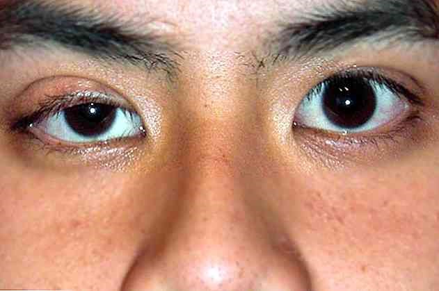
Horner’s syndrome is characterised by ptosis, miosis and anhydrosis due to dysfunction of cervical sympathetic output
Horner’s syndrome occurs when there is a disruption of the cervical sympathetic pathways running from the hypothalamus to the eye
The causative lesion may be primary (including congenital) or secondary to another disorder.
Lesions are generally divided into the following:
- Central (e.g., brainstem ischaemia, syringomyelia, brain tumours)
- Peripheral (e.g. Pancoast tumour, cervical adenopathy, head and neck trauma, aortic or carotid dissection and thoracic aortic aneurysm).
Peripheral lesions may be preganglionic or postganglionic in origin.
Symptomatology of Horner’s syndrome
The symptoms of Horner’s syndrome include ptosis, miosis, anhydrosis and hyperemia of the affected side.
In the congenital form, the iris is not pigmented and remains blue-grey.
Diagnosis of Horner’s syndrome
- Cocaine droplet instillation test
- MRI or CT scan to diagnose the cause
Instillation of eye drops can help confirm and characterise Horner’s syndrome
Initially, drops of cocaine (4-5%) or apraclonidine (0.5%) are placed in both eyes:
- Cocaine: cocaine blocks the synaptic reuptake of noradrenaline and causes dilation of the pupil in the unaffected eye.
- If a postganglionic lesion is present (peripheral Horner’s syndrome), the pupil of the affected eye does not dilate because the postganglionic nerve terminals are degenerated; the result is increased anisocoria.
- If the lesion is above the superior cervical ganglion (preganglionic or central Horner’s syndrome) and the postganglionic fibres are intact, the pupil of the affected eye also dilates and anisocoria decreases.
- Apraclonidine: apraclonidine is a weak alpha-adrenergic agonist that narrows the pupil of a normal eye. If there is a postganglionic lesion (peripheral Horner’s syndrome), the pupil of the affected eye dilates much more than that of the unaffected eye because the iris dilator muscle of the affected eye has lost its sympathetic innervation and developed an adrenergic hypersensitivity. Consequently, anisocoria decreases. (However, the results may be false negative if the injury is acute.) If the injury is preganglionic (or in the case of a central Horner syndrome), the pupil of the affected eye does not dilate because the iris dilator muscle does not develop increased adrenergic sensitivity; consequently, anisocoria increases.
If the results suggest Horner’s syndrome, hydroxyamphetamine (1%) can be inserted into both eyes 48 h afterwards to help localise the lesion.
Hydroxyamphetamine works by causing norepinephrine to be released from presynaptic terminals.

It has no effect if postganglionic lesions are present as these lesions cause degeneration of the postganglionic terminals.
Therefore, when hydroxyamphetamine is applied, the following occurs:
- Postganglionic lesion: the pupil of the affected eye does not dilate, but the pupil of the healthy eye dilates, resulting in increased anisocoria.
- Central or preganglionic lesion: the pupil of the affected eye dilates normally or more than normally, and the pupil of the unaffected eye dilates normally, resulting in decreased or unchanged anisocoria. (However, postganglionic lesions sometimes give the same results).
The hydroxyamphetamine test is performed less frequently than apraclonidine tests, partly because hydroxyamphetamine tends to be available less often.
For results to be valid, the hydroxyamphetamine test should be delayed until 24 h after apraclonidine instillation.
Patients with Horner’s syndrome should undergo MRI or CT of the brain, cord, chest or neck (depending on clinical suspicion) to localise the abnormality.
Treatment of Horner’s syndrome
- Treatment of the cause
If identifiable, the cause is treated; there is no treatment for primary Horner’s syndrome.
Read Also:
Emergency Live Even More…Live: Download The New Free App Of Your Newspaper For IOS And Android
Autoimmune Diseases: The Sand In The Eyes Of Sjögren’s Syndrome
Corneal Abrasions And Foreign Bodies In The Eye: What To Do? Diagnosis And Treatment
Eye Burns: What They Are, How To Treat Them