
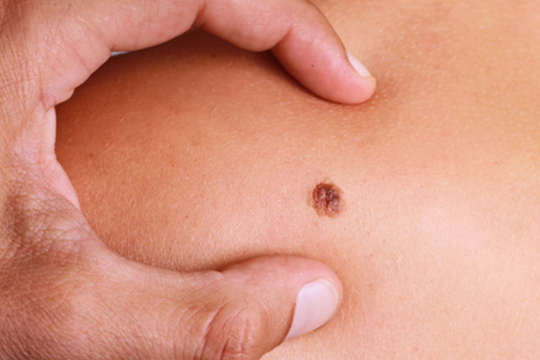
Moles: knowing them to recognise melanoma
We see moles every day, but we often forget that melanoma, one of the most aggressive skin cancers, can look very similar – at least to the naked eye
In fact, in its early stage melanoma can be indistinguishable from a nevus, but the dermatologist can recognise it with the dermatoscope.
What are moles?
Moles are benign tumours of the skin, which arise from the cells responsible for the production of melanin (melanocytes) and vary in colour, shape and size: they can be larger or smaller, light or dark, they can be flat or protrude from the skin.
In some people, a large number of moles (more than 100) can be a sign of a tendency to poorly “control” melanocyte proliferation, and therefore the risk of melanoma.
Some moles may be present at birth, but most appear in childhood and it is normal for their number and size to increase over the years and in a harmonious manner with development, until young adulthood.
From this moment on, the appearance of new “moles” should be brought to the attention of the dermatologist.
But why is it said that it is important to notice if a mole changes?
Let’s dispel a myth: only 30% of melanomas develop from an existing nevus.
The majority of melanomas (about 70-75%, depending on the different studies) develop on healthy skin in the absence of a pre-existing nevus.
However, during the initial, relatively slow phase of its progression, the melanoma already shows (microscopic) characteristics that distinguish it from a benign nevus.
These can be recognised by the dermatologist, provided a person adheres to regular preventive check-ups.
If not removed at this stage, the melanoma grows showing those alterations (in colour, shape, etc.) in the presence of which it is necessary to promptly consult the specialist.
In the last decades the incidence of melanoma in the Italian population has increased, but if it is recognised when it is still at an early stage, almost all patients can achieve definitive cure with a simple outpatient operation under local anaesthesia.
If, on the other hand, timely action is not taken, the risk of mortality unfortunately increases: in fact melanoma, in its progression, acquires metastatic potential.

The main cause of melanoma is excessive exposure to the ultraviolet rays of the sun (from this point of view, it is essential to wear protective cream) and tanning lamps, and it is advisable for those who have had such exposure to see a dermatologist regularly.
Checking moles: a quick and essential method of prevention
For the early detection of melanoma it is good to follow a regular course of prevention, which goes beyond the daily observation of one’s own skin.
All adults should have an annual dermatological examination, but this recommendation is imperative for those with more than a hundred moles, for those who have been exposed to ultraviolet radiation, and for those with phototypes I or II, i.e. particularly fair skin, blue or green eyes and blond or red hair.
People who suffered from sunburn in childhood, first-degree relatives with melanoma or who have already had skin cancer also need to be more careful.
During the examination, the dermatologist examines all moles with a dermatoscope, a small instrument that allows the microscopic characteristics of the mole to be observed more accurately than with the naked eye.
With dermoscopy (or epiluminescence microscopy) it is possible on the one hand to recognise melanoma before it presents the typical features visible to the naked eye, and when it can be cured with a simple surgical removal, and on the other hand to avoid the unnecessary removal of lesions that are apparently alarming on inspection with the naked eye, but on dermatoscopic examination prove to be harmless.
The dermatologist will then assess on a case-by-case basis whether closer examinations are necessary or whether it is necessary to focus on certain moles rather than all the others.
Digital videodermoscopy (so-called mole mapping) allows, through the archiving of digital images, to monitor over time the dermoscopic characteristics of suspicious nevi, making it easier to identify those early melanomas that could escape even dermoscopic examination, for which the evolution in the short term (4-6 months) is an important clue to the diagnosis.
By taking photographs of the entire body surface, at the same time digital videodermoscopy makes it possible to identify with greater certainty nevi that appear where there was no evidence of them at previous checks, and on which it might be appropriate to pay more attention.
Read Also:
What Is A Tumour And How It Forms
Rare Diseases: New Hope For Erdheim-Chester Disease
How To Recognise And Treat Melanoma