
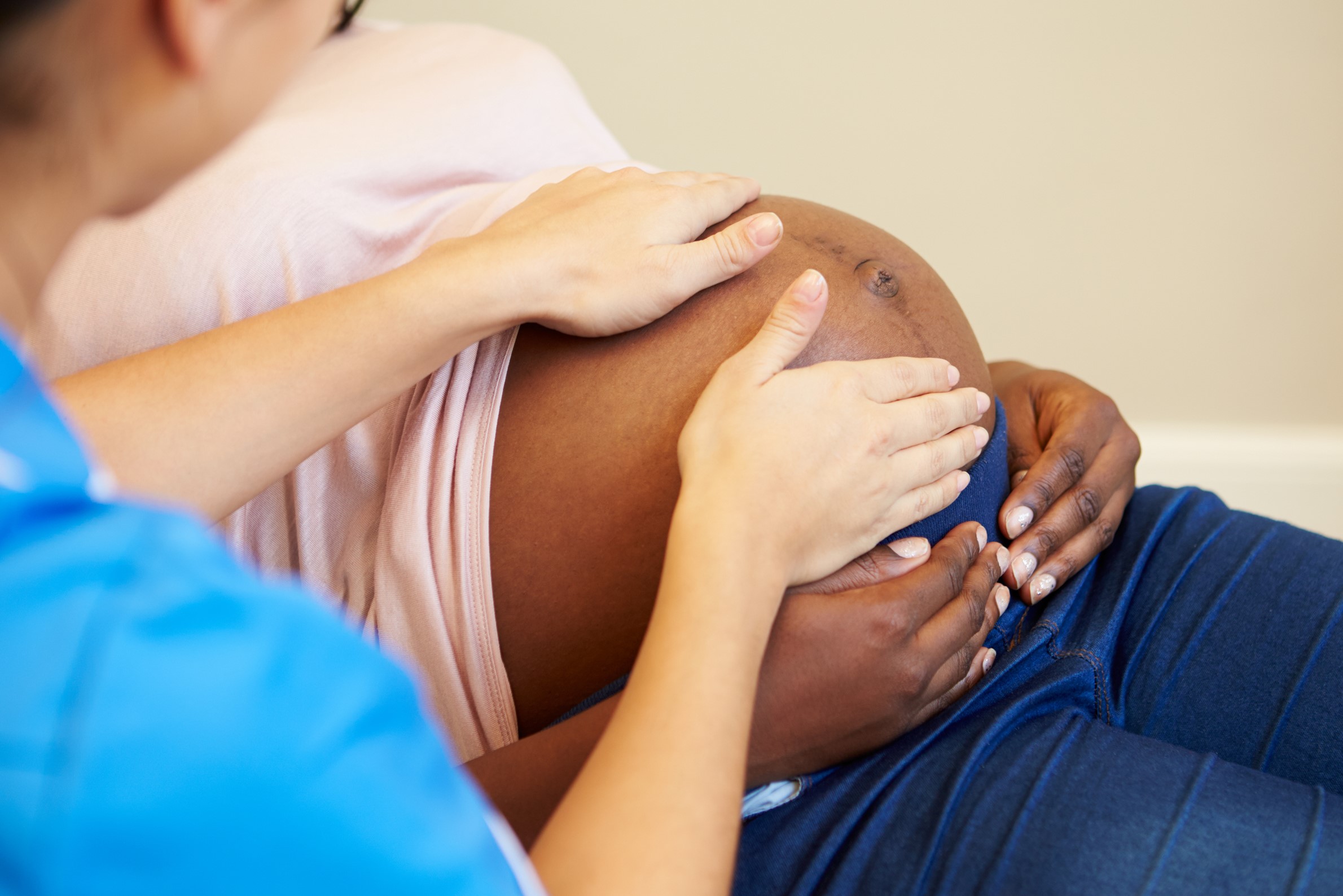
Pregnancy: what it is and when structural ultrasound is necessary
Let’s talk about structural ultrasound: the use of ultrasound has found great impact in obstetrics and gynaecology practice, so much so that all too often the expectations attributed to this technique exceed the real possibilities
Ultrasound, when applied rationally and in expert hands, can give precise and useful, sometimes vital, information.
How ultrasound works
Ultrasound uses ultrasound, intermittent high-frequency waves generated by applying alternating current to a transducer made of piezoelectric material.
It has the ability to send ultrasound waves and receive reflected waves from the underlying tissue.
The response is then processed by the computer, which gives us the characteristic two-dimensional greyscale image.
Use and interpretation of the ultrasound test
Ultrasound has been used extensively in recent years because it has the potential to identify foetal malformations.
Should ultrasound be given to all pregnant women or only to those at particular risk? Routine use of ultrasound is more expensive and less accurate because it is quicker.
On the other hand, selective ultrasound testing requires more time and highly specialised personnel.
Certainly, all pregnant women should undergo at least one ultrasound test.
The recent guidelines of the Italian Society of Obstetrical-Gynaecological Sonography suggest at least two ultrasound examinations, one at the beginning of gestation and one around 20 weeks, the so-called structural ultrasound and further tests only if necessary.
What can be seen with structural ultrasound
This test should be carried out between 20 and 22 weeks, when the development of internal organs is sufficiently advanced to be able to define their completeness and normality with good approximation, while the bones are still mainly in the cartilaginous state and are still being passed through by ultrasound.
Structural ultrasound allows an estimate of fetal growth, in relation to weeks of gestation.
Knowing the weight at this stage is of no use because it will change in the following months.
The test of the skull distinguishes the brain, cerebellum, corpus callosum and some minor structures that are important for ruling out major abnormalities of the central nervous system, such as hydrocephaly or anencephaly.
Of the face, the orbital cavities, mouth, upper lip in anatomical continuity with the hard palate can be visualised to rule out cleft lip formation.
Sometimes dental buds are also visible.
The test of the chest gives us the exact position of the heart and the absence of cystic malformation of the lung.
Of the heart we must see the four chambers, two atria and two ventricles, and the two axes, i.e. the continuity between the cardiac cavity and the great vessels.
Of the abdomen we distinguish the abdominal wall, which should be completely closed except for the point where the umbilical cord originates; the stomach, the liver, the gallbladder, the two kidneys, the bladder are structures that are fairly easy to identify.
Of the skeleton, in addition to the diameters of the head, the length of the femur and humerus are to be measured; both segments (proximal and distal, i.e. arm and forearm, leg and thigh) for each of the four limbs, and the hands and feet are to be visualised, while the fingers are not to be counted.
The spine should be studied in longitudinal and transverse section, especially the sacral segments, for the diagnosis of spina bifida.
It is always useful to indicate the position of the placenta and the amount of amniotic fluid.
Limitations of structural ultrasound
It is often difficult to visualise the upper lip and hard palate, the face, the development of the heart ventricles, the genitals and some peripheral vessels.
If the position of the foetus prevents the heart from being tested, the ultrasound scan should be repeated half an hour later or the following day.
Some findings, such as the amount of amniotic fluid, low placental insertion or abnormal placental anatomy, are a matter of subjective interpretation because there are no precise methods of measurement.
Some developmental defects may appear after the structural test, for example, hydrocephaly, hydronephrosis, gastrointestinal atresia.
Growth defects may also occur at any time during pregnancy.
Reliability and accuracy depend on the experience of the operator, the echogenicity of the tissues, and the position of the foetus; at present, sonographers who perform the structural scan when faced with a diagnostic doubt invite the patient to go to a centre specialised in the diagnosis of that particular pathology.
Read Also
Emergency Live Even More…Live: Download The New Free App Of Your Newspaper For IOS And Android

Ultrasound: What It Is And When It Is Performed
Hip Dysplasia: The First Ultrasound Scan After 40 Days Of Life
Colposcopy: How To Prepare, How It Is Performed, When It Is Important
Breast Cancer: For Every Woman And Every Age, The Right Prevention
Transvaginal Ultrasound: How It Works And Why It Is Important
Pap Test, Or Pap Smear: What It Is And When To Do It
Mammography: A “Life-Saving” Examination: What Is It?
Breast Cancer: Oncoplasty And New Surgical Techniques
Gynaecological Cancers: What To Know To Prevent Them
Ovarian Cancer: Symptoms, Causes And Treatment
What Is Digital Mammography And What Advantages It Has
What Are The Risk Factors For Breast Cancer?
Breast Cancer Women ‘Not Offered Fertility Advice’
Abnormal Uterine Bleeding: Causes And Tests To Be Performed
How Does Cystitis Manifest Itself?
Cystitis, Antibiotics Are Not Always Necessary: We Discover Non-Antibiotic Prophylaxis
Vulvodynia: What Are The Symptoms And How To Treat It
What Is Vulvodynia? Symptoms, Diagnosis And Treatment: Talk To The Expert
Accumulation Of Fluid In The Peritoneal Cavity: Possible Causes And Symptoms Of Ascites
What’s Causing Your Abdominal Pain And How To Treat It
Pelvic Varicocele: What It Is And How To Recognise The Symptoms
Can Endometriosis Cause Infertility?
Candida Albicans And Other Forms Of Vaginitis: Symptoms, Causes And Treatment
What Is Vulvovaginitis? Symptoms, Diagnosis And Treatment
Vaginal Infections: What Are The Symptoms?
Chlamydia: What Are The Symptoms And How To Treat It
Chlamydia, Symptoms And Prevention Of A Silent And Dangerous Infection
Spotting, Or Atypical Female Bleeding: What It Is And The Diagnostic Pathway
What Are The Symptoms Of Urethritis And How Is It Treated?