
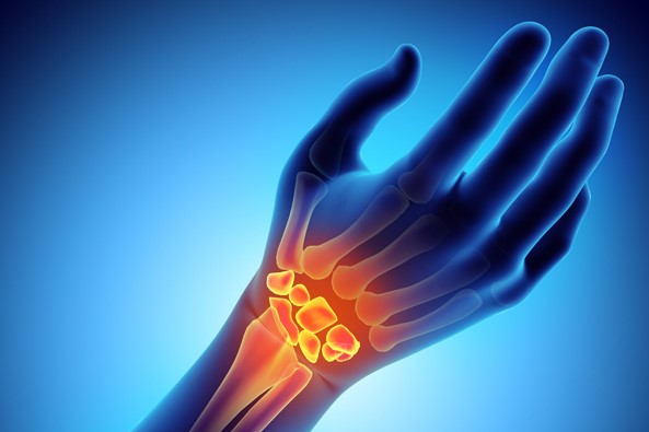
Wrist fracture: how to recognise and treat it
A wrist fracture is a common occurrence, both in the work of a rescuer and in the daily life of an ordinary citizen, so recognising it is essential
The causes of wrist fracture
The causes of wrist fractures are mainly to be found in traumas.
In the case of sportsmen and women, there are certain specific sports which, due to their particular characteristics, are more prone to this problem.
These include:
- contact sports such as boxing and martial arts;
- sports with a greater risk of falling, such as volleyball, football, basketball, tennis;
- speed sports, e.g. running, motorcycling, cycling;
- winter sports, e.g. skiing, skating, snowboarding.
Falls are one of the main causes of wrist fractures because, when falling, people tend to bring their hands forward to protect their head instinctively, in some cases severely compromising the wrist joint.
Other causes associated with wrist fracture are accidents (domestic, road), but also osteoporosis, a condition of bone degeneration, which is very common in the elderly and in menopausal women.
Types of wrist fracture
Among the different fractures that can affect the wrist, the most frequent are:
- fracture of the scaphoid
- fracture of the distal epiphysis of the radius and ulna.
Wrist fractures: Scaphoid fracture
A fracture of the scaphoid is painless, or at least it hurts for a few days, without suggesting the presence of a fracture.
Very often, the person does not even consult a specialist and can live with it for years before discovering it.
For vascular reasons, this type of fracture heals with extreme difficulty, as it is a bone that is poorly vascularised (i.e. supplied with blood vessels).
If it is not treated, a situation of pseudo-arthrosis is created, i.e. an unhealed fracture, which in turn gives rise to a condition of mechanical instability of the wrist joint, the antechamber to arthrosis proper.
Because of the shape of the scaphoid, a spongy bone with an extremely complex shape, the fracture is sometimes not visible, despite the X-rays taken in the emergency room.
For this reason it is necessary to take special care in accurately assessing the fracture, subjecting the patient to a CT scan or further X-rays at a distance of 1 week / 10 days.
Fractures of the distal epiphysis of the radius and fracture of the radius and ulna
Fractures of the distal epiphysis of the radius and fractures of the radius and ulna are the most common.
For wrist fractures, today, there is a very strong re-evaluation and difference in approach between a more traditional orthopaedic surgeon and a specialist hand surgeon.
Very often fractures indicated for surgery are treated with plaster casts in unnatural positions in an often unsuccessful attempt to keep an unstable fracture reduced, resulting in an unsatisfactory aesthetic and functional outcome.
A very high percentage of wrist fractures treated with plaster casts undergo secondary decomposition which can be more or less severe.

Hand surgeons see many of these patients and often have to operate on wrist fractures that are in poor condition because they may have been in plaster for weeks before they realised that they needed immediate surgery.
Treating them surgically when they are already almost firmly established becomes much more difficult, so it is always better to rely on a hand specialist who can recognise them and initiate the most suitable diagnostic and therapeutic procedure.
Wrist fracture: diagnosis and treatment
In addition to the clinical evaluation by the hand specialist and conventional X-rays, another examination that can provide a definitive diagnosis is undoubtedly the CT scan.
Treatment for wrist fracture is often surgical
In more than 50% of cases, a wrist fracture is associated with a ligamentous injury, which often cannot be diagnosed in the acute phase due to the inability to clearly investigate the involved part.
How the surgery works
The operation consists of an osteosynthesis with the insertion of a plate, which is able to return the fracture fragments to their original position.
Compared to a few years ago, the means of synthesis have undergone a great evolution: we no longer have only one type of plate, but a wide range of plates to meet the needs of different types of fracture.
These means of synthesis are also perfectly efficient because they do not interfere with or obstruct the correct movement of the tendons, and thanks to the use of screws that block both the bone and the plate, they guarantee greater stability.
When we operate on wrist fractures, once the radius plate is in place, we always perform an X-ray assessment to detect any gross ligament injuries, which are immediately treated. Even in the case of injured ligaments, we proceed surgically through the use of means of synthesis (e.g. small anchors or wires), which can hold the bones together so that the ligament can reattach.
Surgery for a broken wrist
In cases of very fragmented fractures, a two-pronged approach is chosen:
- traditional: to insert the plate;
- arthroscopic: to reduce the small fragments in the joint.
In sportsmen and women, in order to ensure the quickest possible resumption of competitive activity and a return to the field, it is often necessary to opt for aggressive therapeutic choices in order to reduce the time of inactivity.
In the post-surgery period, there are generally several options:
- a specific brace that the patient has to remove several times a day for exercise;
- a splint to be worn for 5 weeks with a subsequent X-ray.
If, after the X-ray, the fracture is consolidated, the rehabilitation can begin.
Read Also:
Spencer WOW, What’s Going To Change In Patient Transport?