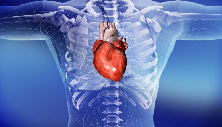
Cardiac tamponade: symptoms, ECG, paradoxical pulse, guidelines
In medicine, ‘cardiac tamponade’ refers to an abnormal accumulation of fluid or blood within the pericardial cavity that leads to alterations in the cardiovascular system
Cardiac tamponade can be acute or chronic and is characterised by a series of linked haemodynamic events that can progress to cardiovascular collapse
Myocardial free wall rupture tamponade is most often present in the elderly with a cardiac history, such as a previous acute myocardial infarction.
Pathophysiology
Accumulation of blood in the pericardial cavity, which is normally a virtual cavity, results in:
- an increase in intrapericardial pressure;
- the increase in pressure causes a rise in central venous pressure, which is intended to maintain cardiac filling and prevent ventricular wall collapse;
- this leads to a reduction in venous return to the heart;
- at the same time the transmural pressure (i.e. diastolic pressure minus pericardial pressure) is reduced to zero leading to a reduction in preload.
The end result is therefore a rise in atrial and pericardial pressure, an inspiratory reduction in systolic blood pressure (paradoxical pulse) and arterial hypotension.
Causes of pericardial tamponade
In a healthy pericardium there is between 25 and 50 ml of fluid, known as pericardial fluid, which serves to lubricate and reduce the friction that would occur during the mutual sliding of the two pericardial leaflets.
As the fluid increases, the pericardial pressure will increase proportionally and we will have different clinical and symptomatic pictures: if the fluid increases suddenly as in the case of a rupture of the myocardial wall, the intrapericardial pressure increases rapidly and can exceed the intracardiac pressure, leading to cardiac tamponade.
Several studies have shown that symptoms can already occur at around 100 ml.
The causes that can lead to such an acceleration are:
- venous and lymphatic obstruction, following intracardiac manoeuvres, such as pacemaker insertion, or during cardiac catheterisation
- tumour and/or metastases;
- trauma affecting the myocardium, e.g. in a car or sports accident.
Various diseases can lead to the development of tamponade, including neoplasms and chronic inflammatory diseases of the pericardium: however, the course is less rapid and heart failure with peripheral oedemas will manifest itself over a longer period of time.
The main causes of cardiac tamponade are:
1) blood collections secondary to:
- penetrating wounds or severe blunt trauma;
- rupture of aortic or coronary aneurysms;
- rupture of the heart in the course of acute myocardial infarction;
- myocardial perforation during cardiac catheterisation, pacemaker placement, sternal bone marrow biopsy, pericardiocentesis;
- haemorrhagic diathesis or treatment) anticoagulant (haemorrhagic exudative collections).
2) serous or exudative collections secondary to:
- acute pericarditis of viral, bacterial, tuberculous, neoplastic, uremic aetiology;
- cardiac and extracardiac neoplastic processes (paraneoplastic syndromes);
- anasarca.
Symptoms of cardiac tamponade
Mild cardiac tamponade may be asymptomatic, whereas moderate and severe forms manifest themselves with symptoms such as dyspnoea, angina pectoris and dizziness.
The presence of the so-called paradoxical pulse, i.e. the reduction of arterial pressure in inspiration beyond the physiological 10 mmHg, together with the increase in venous pressure, visible as jugular turgor, arterial hypotension and the perception of muffled heart tones (Beck’s triad), often leads to the absence of peripheral pulse perception, even in the presence of normal electrical activity (electromechanical dissociation).
Schematically, the symptoms of tamponade are:
- reduction in systolic pressure during the inspiratory phase;
- sense of precordial (chest) pain and oppression;
- dyspnoea;
- tachycardia (increased heart rate);
- arterial hypotension (lowering of blood pressure);
- distant and muffled tones;
- paradoxical Kussmaul pulse (reduction in pulse amplitude, until disappearance, during the inspiratory phase);
- Kussmaul’s sign (inspiratory distension of the neck veins);
- turgor of the veins of the neck and upper limbs, secondary to increased venous pressure;
- shock.
Diagnosis of cardiac tamponade
The diagnosis of cardiac tamponade is suspected through the clinic (history and objective examination) and confirmed by means of
- electrocardiogram.
- chest X-ray: shows enlargement of the cardiac shadow with uncongested pulmonary fields
- echocardiography: during cardiac tamponade, the velocity of the tricuspid and pulmonary flows increases with inspiration, while that of the aortic and mitral flows decreases, since this is observed in almost all cases, the absence of this element suggests the presence of a non-“tamponade” effusion
- cardiac catheterisation: this is useful if one wants to be certain of the diagnosis in doubtful cases, by measuring the right atrial pressure, which in the course of tamponade is equal to the pericardial pressure, whereas it is normally higher.
ECG in cardiac tamponade
On the electrocardiogram performed on a patient with cardiac tamponade, one notices
- inconstant electrical alternation of the QRS and P and T waves;
- decreased voltage of the P wave, QRS (in no peripheral lead is the R wave higher than 5 mm and in no precordial lead is the R wave higher than 10 mm) and T wave.
ECG EQUIPMENT? VISIT THE ZOLL BOOTH AT EMERGENCY EXPO
The differential diagnosis of cardiac tamponade must be made primarily with:
- cardiogenic shock;
- acute right congestive decompensation.
In both cases, a paradoxical pulse is uncommon.
Paradoxical pulse: how is it assessed?
A paradoxical pulse is a significant decrease in pulse amplitude and systolic pressure of more than 10 mmHg during inspiration.
A slight reduction in systolic pressure secondary to a relative increase in blood in the pulmonary vessels during inspiration is normal, whereas in tamponade the reduction is more pronounced.
The magnitude of the paradoxical pulse can be quantified with a sphygmomanometer: it is equal to the difference in pressure audible on exhalation at the first Korotkoff tone and the pressure level at which the tones are audible in all phases of the respiratory cycle.
The inverted form (decrease in systolic and diastolic pressure during exercise), on the other hand, is an indication of hypertrophic cardiomyopathy.
Treatment of cardiac tamponade
The treatment protocol includes:
- the prompt admission of the patient, possibly to an intensive care unit, in order to perform pericardiocentesis and possibly pericardiectomy;
- the avoidance of reducing venous pressure with bloodletting and diuretics since venous hypertension, by balancing the increase in intrapericardial pressure, ensures a certain degree of cardiac filling, representing a temporary compensation mechanism.
It is important to remember that the removal of even small amounts of fluid (even below 100 ml) rapidly leads to a good improvement in symptoms and haemodynamics, as it changes the pericardial pressure/volume ratio, which is why drainage is the most important therapy in the patient with cardiac tamponade.
Pericardiocentesis
When tamponade is low pressure (less than 10 cm of water), pericardiocentesis is preferred not to be used.
On the contrary, in more severe cases, the drainage procedure must be used: surgically (via subxiphoid incision or video-assisted thoracoscopy) or percutaneously, with a needle or balloon catheter.
The advantages of ‘covered’ needle drainage are related to the echo-guided method: the simplicity of inserting and leaving the catheter in place even for days and being able to administer drugs directly into the pericardial space.
The lesser trauma and the possibility of haemodynamically following the drainage, guide the timing for removal, which is generally discouraged unless the residual fluid is around 25 ml.
The advantages of ‘open’ drainage on the operating table are related to the possibility of complete removal of fluid, direct access to tissue for possible biopsies and the possibility of draining localised effusions.
Read Also:
Emergency Live Even More…Live: Download The New Free App Of Your Newspaper For IOS And Android
Cardiomyopathies: What They Are And What Are The Treatments
Heart Disease: What Is Cardiomyopathy?
Inflammations Of The Heart: Myocarditis, Infective Endocarditis And Pericarditis
Heart Murmurs: What It Is And When To Be Concerned
Broken Heart Syndrome Is On The Rise: We Know Takotsubo Cardiomyopathy
What Is A Cardioverter? Implantable Defibrillator Overview


