
Chest trauma: clinical aspects, therapy, airway and ventilatory assistance
Trauma is currently one of the most serious public health problems worldwide: in industrialised countries, it is the leading cause of death in the under-40 age group and the third leading cause of death after heart disease and cancer
In about a quarter of cases, injuries lead to a disability that requires the patient to be bedridden and undergo complex treatment and a period of rehabilitation.
Given the young age of most of these patients, trauma is responsible – economically speaking – for more severe disability and loss of productivity overall than even heart disease and cancer taken together.
Clinical aspects of chest trauma
An accurate history of the manner and circumstances of the trauma is vital for the assessment of the extent of the injury sustained.
It is important, for example, to gather information about the manner of the motor vehicle accident (were the seat belts fastened?, was the victim thrown from the passenger compartment?, what were the dimensions of the vehicle?, and so on), the calibre and type of weapon used, the time elapsed before help arrived, whether there was any shock at that stage.
Pre-existing cardiac, pulmonary, vascular or renal diseases, or drug or alcohol abuse, can also influence the body’s reaction to trauma.
A quick but careful objective examination should be conducted to assess airway patency, breathing patterns, blood pressure, the presence of signs of flail chest or subcutaneous emphysema, symmetry and other features of the pulmonary auscultatory findings.
A rapid and systematic approach for the initial assessment of the nervous, circulatory and respiratory systems is a simple point rating system for the severity of the trauma patient’s clinical condition.
This trauma score takes into account the Glasgow coma scale, maximum arterial pressure and breathing rate: the three parameters are given a score from zero to four, where four indicates the best condition and zero the worst.
Finally, the three values are added together.
Let’s take an example of a patient with:
Glasgow coma scale: 14;
blood pressure: 80 mmHg;
respiratory rate = 35 breaths per minute.
Trauma score = 10
We remind the reader that the Glasgow coma scale is a neurological assessment system, which scores according to the best ocular, verbal and motor responses to various stimuli.
In a study of 2166 patients, a modified ‘trauma score’ was shown to discriminate patients who would survive from those who were fatally injured (e.g. scores of 12 and 6 were associated with 99.5% and 63% survival, respectively), allowing for more rational triage to the various trauma centres.
Based on these initial assessments, the subsequent diagnostic and therapeutic protocol is decided.
Numerous instrumental and laboratory tests are frequently used to better define the nature and extent of the thoracic injuries reported. An anteroposterior (AP) X-ray is virtually always necessary for further evaluation of the patient and as a guide to emergency treatment.
Complete blood count (CBC), electrolyte assay, arterial blood gas analysis (ABG) and electrocardiogram (ECG) are performed on admission and then serially.
More sophisticated investigations such as CT, magnetic resonance imaging (MRI) and angiography help to define the extent and severity of injuries more precisely.
Treatment of chest trauma
Approximately 80% of all trauma-related deaths occur in the first few hours after the event.
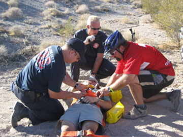
Survival is dependent on the rapid activation of life-support procedures and transport to a trauma centre.
Immediate treatment of chest trauma victims involves maintaining airway patency, oxygen therapy with an FiO of 1.0 (e.g., with a ‘non-rebreathing’ mask, ‘balloon’ ventilator or high-flow oxygen delivery equipment) mechanical ventilation, placement of peripheral and central intravenous (EV) lines for administration of fluids and blood, application of a chest drain, and possibly immediate transfer to the operating room (OR) for an emergency thoracotomy.
The introduction of a pulmonary artery catheter is useful for the treatment of patients who are haemodynamically unstable and/or require large fluid infusions to maintain electrolyte balance.
The treatment of pain is also important.
The use of patient-controlled analgesic (PCA) dispensers (e.g. systemic infusion or thoracic epidural) improves pain tolerance, deep breathing cooperation, lung function and makes the need for ventilatory assistance less frequent.
Airway assistance
Airway obstruction is generally considered the most important correctable cause of death in trauma patients.
This condition is most often caused by the tongue sliding backwards into the oropharynx.
Aspiration of vomit, blood, saliva, dentures, and oedema following an oropharyngeal injury are alternative causes of airway obstruction.
Placing the patient’s head in a suitable position and inserting an oropharyngeal cannula helps to maintain airway patency and allows 100% oxygen to be delivered with a balloon mask.
In most emergency cases, the artificial airway of choice is an endotracheal cannula of appropriate calibre, with a sleeve, which allows positive pressure ventilation, facilitates endotracheal suction and helps protect the lung from aspiration of gastric contents.
If a cervical fracture is suspected, the insertion, under bronchoscopic control, of a nasotracheal cannula is recommended, because this procedure requires less extension of the head.
Manoeuvres for the placement of the endotracheal cannula can trigger cardiac arrest, mediated by inadequate pre-oxygenation, intubation of a main bronchus or oesophagus, respiratory alkalosis secondary to over-intensive ventilation, and/or a vasovagal reflex.
Careful monitoring of correct cannula placement is necessary to ensure that both lungs are ventilated.
Indeed, in approximately 30% of patients undergoing resuscitation manoeuvres, intubation of the right main bronchus occurs.
A chest X-ray and a fibrobronchoscopy allow the detection of blood accumulations, which need to be aspirated.
A fibreoptic bronchoscopy, either diagnostic or therapeutic, often proves very useful in patients with persistent or recurrent atelectasis.

In patients with severe asymmetric lung contusions or tracheobronchial ruptures, who require independent lung ventilation, the use of a double-lumen tracheal cannula may be necessary.
If endotracheal intubation or placement of a tracheostomy cannula is difficult or impractical, a cricothyrotomy may be performed until tracheostomy can be performed.
In the absence of other feasible accesses, the introduction of a 12-gauge needle by the cricothyroid route may allow, in the short term, percutaneous transtracheal ventilation and oxygenation, pending the placement of a tracheostomy cannula.
Ventilatory care
Patients who come to observation in apnoea, in impending respiratory failure (respiratory rate above 35/minute), or in full respiratory failure (PaO2 below 60 mmHg, PaCO2 above 50 mmHg, and pH below 7.20) require respiratory assistance.
The parameters of ventilatory assistance for a patient with thoracic injuries of unknown severity should be set to provide full support by means of volume-dependent assistance-control ventilation, with a tidal volume of 10 ml/kg, a rate of 15 cycles/minute, an airflow rate to ensure an inspiration/exhalation (I:E) ratio of 1:3, and a FiO2 of 1.0.
These parameters can be changed after a more thorough clinical examination and once ABG results are available.
Frequently, a PEEP of 5-15 cm Hp is necessary to improve lung volume and oxygenation.
However, the use of positive pressure ventilation and PEEP in patients with chest trauma requires extreme caution, in relation to the risk of inducing hypotension and barotrauma.
Once the patient has regained the ability to breathe spontaneously more efficiently, intermittent, synchronised forced ventilation (IMSV), combined with pressure support (PS), facilitates weaning from the ventilator.
The last step before extubation is to check the patient’s spontaneous breathing capacity with continuous positive pressure (CPAP) at 5 cm H2O to maintain adequate oxygenation and improve lung mechanics.
In complicated cases, numerous, more complex alternative ventilation and gas exchange support systems may be used.
In severe forms ofARDS, the use of pressure-dependent, inverse-ratio ventilation can improve ventilation and oxygenation and help reduce peak airway pressure.
Patients with severe asymmetric lung injury who experience hypo-oxygenation during conventional mechanical ventilation, despite PEEP and 100% oxygen delivery, may benefit from independent lung ventilation using a double-lumen tracheal cannula.
Independent lung ventilation or high-frequency ‘jet’ ventilation can meet the needs of patients with bronchopleural fistula.
In adults, extracorporeal membrane oxygenation (ECMO) is apparently no more effective than conventional mechanical ventilation.
ECMO seems, on the other hand, preferable in the paediatric population.
Once multiple organ failure secondary to trauma has been corrected, ECMO may also be more effective in adults.
Other respiratory assistance techniques
The thoracic trauma patient often requires additional forms of treatment.
Airway humidification, with heated or unheated vapours, is frequently practised to control secretions.
Airway hygiene is also essential in intubated subjects or those with mucus retention.
Respiratory physiotherapy is often useful for the mobilisation of secretions retained in the airways and can help re-expand atelectasis areas.
Frequently, bronchodilators in the form of aerosols are used to reduce airway resistance, facilitate lung expansion and reduce respiratory work.
These forms of ‘low-tech’ respiratory care are all very important in the management of the thoracic trauma patient.
Read Also:
Emergency Live Even More…Live: Download The New Free App Of Your Newspaper For IOS And Android
Tracheal Intubation: When, How And Why To Create An Artificial Airway For The Patient
What Is Transient Tachypnoea Of The Newborn, Or Neonatal Wet Lung Syndrome?
Traumatic Pneumothorax: Symptoms, Diagnosis And Treatment
Diagnosis Of Tension Pneumothorax In The Field: Suction Or Blowing?
Pneumothorax And Pneumomediastinum: Rescuing The Patient With Pulmonary Barotrauma
ABC, ABCD And ABCDE Rule In Emergency Medicine: What The Rescuer Must Do
Multiple Rib Fracture, Flail Chest (Rib Volet) And Pneumothorax: An Overview
Internal Haemorrhage: Definition, Causes, Symptoms, Diagnosis, Severity, Treatment
Cervical Collar In Trauma Patients In Emergency Medicine: When To Use It, Why It Is Important
KED Extrication Device For Trauma Extraction: What It Is And How To Use It
How Is Triage Carried Out In The Emergency Department? The START And CESIRA Methods