
CT, MRI and PET scans: what are they for?
CT, MRI and PET scans are three widely used diagnostic tests. But how do they differ and when should one or the other be used?
We have different examinations because none of them can be considered the best of all: each examination has the ability to see our bodies in a different way.
What are CT, MRI and PET scans?
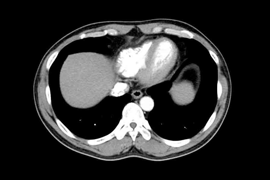
CT (Computed Axial Tomography) is perhaps the best known examination and through the use of X-rays allows us to observe differences in density and shape of organs.
Magnetic resonance imaging (MRI or NMR) provides an idea of how water molecules are distributed in our bodies.
PET (Positron Emission Tomography) uses specific substances, called radiopharmaceuticals, to observe particular structures or organs and is able to recognise certain metabolic processes.
A CT scan therefore gives us an idea of the shape, MRI not only gives us an idea of the shape but also adds a functional component, while PET gives us an idea of the function: a tissue may appear to be more functional because it consumes more energy (i.e. more sugar) and could therefore be cancerous, or it has certain metabolic characteristics that can be defined using radiopharmaceuticals.
PET and the different radiopharmaceuticals
PET does not exist as a single method because different things can be observed depending on the radiopharmaceutical used.
For example, the brain is the organ in our body that consumes the most sugar, and if there are areas that function less we can observe a lowering of sugar consumption.
How do we notice this? We inject a substance that is taken up by the cells as if it were sugar and that has a bulb attached to it that emits positrons.
But we can also see how the blood flow in the brain is, for example by injecting water that has been made radioactive, or observe amyloid deposits, which may be increased in patients with Alzheimer’s disease.

Whereas with CT (even with contrast medium) we only have a density image and with MRI we see soft tissue better because it is rich in water, with PET we have more possibilities depending on the radiopharmaceutical used: sugar consumption, amyloid deposits, bone remodelling or the expression of particular receptors in certain tumours.
PET is therefore an examination that is defined as functional because it allows us to see certain peculiar characteristics of tissues.
Are there examinations capable of merging the different methods, i.e. CT, MRI and PET?
We currently speak of hybrid or multimodal imaging: PET machines perform PET and CT scans because PET is a functional examination and therefore does not provide an idea of the exact anatomy of the lesion or of the alteration that we can localise, while CT is a method that gives us a specific and precise assessment of the anatomy and therefore PET-CT allows us to have the best of both methods.
There are also machines that perform PET and MRI at the same time.
In fact, one imaging method is almost never enough to get all the information we need.
Read Also:
What Is An ECG And When To Do An Electrocardiogram
Inflammations Of The Heart: Myocarditis, Infective Endocarditis And Pericarditis
MRI, Magnetic Resonance Imaging Of The Heart: What Is It And Why Is It Important?