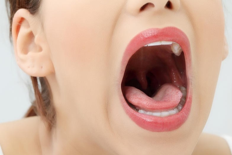
Malignant tumours of the oral cavity: an overview
Malignant tumours (cancer) of the oral cavity are lesions that originate from the uncontrolled proliferation of genetically damaged oral mucosa cells
A large proportion of carcinomas of the oral cavity (15-40%) arise on already known lesions and precancerous conditions (leukoplakia, erythroplasia, lichen, submucosal fibrosis, Fanconi anaemia).
Cancer of the oral cavity may originate in the cheek mucosa, hard palate, anterior part of the tongue, lips, gingival mucosa, retromolar trigone and minor salivary glands.
Malignant tumours of the oral cavity, what are the symptoms?
It can be clinically manifested by the appearance of a granule-like, flat, mammelike or vegetating, whitish or hyperemic, often ulcerated, painful, easily bleeding lesion, which does not heal spontaneously and may cause pain at rest, pain on swallowing and/or chewing, in some cases radiating to the ear, difficulty in swallowing, chewing and speech articulation.
Patients with this disease may progressively feed with increasing difficulty, lose weight and become debilitated.
In other cases, the tumour may manifest directly as a laterocervical lymph node tumefaction, i.e. a mass in the lateral cervical region that is hard to palpate, not very mobile in the underlying planes, with intact skin, of increasing volume, an expression of regional metastatisation.
Who does it affect?
Men were the most likely to develop this tumour but to date the incidence is similar between men and women due to a proportional increase in alcohol and tobacco consumption in women.
The average age of onset is around 50-60 years.
Risk factors predisposing to oral cavity tumours are:
- smoking cigarettes, cigars, pipes and certain types of ‘self-made’ cigarettes; the high concentration of carcinogenic substances contained in tobacco make it very harmful and capable of irreversibly damaging the cells of the oral mucosa;
- alcohol abuse: alcohol drinkers have a 6 times higher risk than non-drinkers.
Their synergistic effect is well known, multiplying the risk of developing oral cancer by as much as 80 times.
In addition to alcoholism and smoking, another important etiopathogenic factor is microtrauma from dental anomalies, poorly preserved or altered dentures or prostheses (frequent in elderly individuals).
There is a small proportion (<5%) of HPV oral cavity carcinomas related to chronic Papilloma Virus infection, a virus with high oncogenic power.
However, it is correct that 25% of oral cancer patients do not drink or smoke.
Oral tumours – the diagnosis
In order to arrive at a diagnosis, it is essential to carry out a thorough anamnestic collection and a thorough otolaryngological objective examination.
Often it is the dentist who sends the patient to the specialist for the detection of suspicious lesions worthy of further investigation.
The biopsy of the lesion is the crucial element for the diagnosis; it is often performed on an outpatient basis after administration of local anaesthesia.
The purpose of the biopsy is to take macroscopically suspicious material that will then be analysed and studied by the anatomo-pathologist.
The most frequent histotype is undoubtedly squamous cell carcinoma in situ or infiltrating.
Treatments
Based on the clinical staging, i.e. the loco-regional and distant extension of the tumour, the case is discussed collegially with oncologist colleagues, radiologists, radiotherapists and anatomo-pathologists in order to propose the best treatment options to the patient.
Surgery is the treatment of choice, especially in tumours of limited size.
Surgery, which is performed by the otorhinolaryngologist (head and neck surgeon), involves radical removal of the tumour, possible reconstruction with flaps taken from other sites, and mono- or bilateral laterocervical lymph node emptying.
Surgical treatment, depending on the final histological examination, may be followed by radiotherapy or concomitant radio-chemotherapy.
What are the results?
Depending on the site and initial extent of the tumour, the overall disease control rate is around 65% with extremes ranging from 95% for small tumours of the lip to 20% for large tumours of the tongue or retromolar trigone.
The probability of loco-regional control varies depending on the presence or absence of lymph node metastases and their extent.
How can this pathology be prevented?
Prevention of these tumours involves abstaining from smoking and alcohol consumption and a screening programme in which the otorhinolaryngologist and dentist are the reference figures.
Read Also:
Emergency Live Even More…Live: Download The New Free App Of Your Newspaper For IOS And Android
Neuroendocrine Tumours: An Overview
Benign Tumours Of The Liver: We Discover Angioma, Focal Nodular Hyperplasia, Adenoma And Cysts
Tumours Of The Colon And Rectum: We Discover Colorectal Cancer
Tumours Of The Adrenal Gland: When The Oncological Component Joins The Endocrine Component
Brain Tumours: Symptoms, Classification, Diagnosis And Treatment
What Is Percutaneous Thermoablation Of Tumours And How Does It Work?
Colorectal Resection: In Which Cases The Removal Of A Colon Tract Is Necessary
Thyroid Cancers: Types, Symptoms, Diagnosis
Tumours Of Endothelial Tissues: Kaposi’s Sarcoma
Gastrointestinal Stromal Tumor (GIST)
Juvenile Gastrointestinal Polyposis: Causes, Symptoms, Diagnosis, Therapy
Diseases Of The Digestive System: Gastrointestinal Stromal Tumours (GISTs)


