
Diseases of the digestive system: gastrointestinal stromal tumours (GISTs)
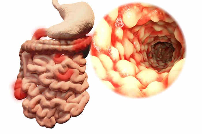
Gastrointestinal stromal tumours (GISTs) are rare tumours of the digestive system that originate from the muscular wall of the oesophagus, stomach, intestine and anal canal
These tumours originate from the interstitial cells of Cajal, smooth muscle cells of the gastrointestinal tract responsible for the physiological contraction movement of the digestive tract.
It is a rare pathology that manifests itself mainly between the ages of 55 and 65, there are rare cases before the age of 40 and only sporadic cases in young children.
It mainly affects the male sex.
At the origin of the onset of GISTs is a mutation in a gene that leads to the uncontrolled proliferation of these cells, which accumulate and give rise to the tumour mass.
Some GISTs (around 5% of cases) are not linked to known genetic alterations, particularly those arising in paediatric age and those associated with syndromes such as neurofibromatosis type 1.
The symptomatology of GISTs may be nuanced, related to disorders of the involved gastrointestinal tract
Diagnostic investigations may include:
- Ultrasound: this is a non-invasive method, with no contraindications, useful in the initial definition of large masses.
- Endoscopy: by means of oesophago-gastro-duodenoscopy (EGDS) or colonoscopy, if lesions of the lower digestive tract are involved, it is possible to visualise a wall bulge, usually lined with regular mucosa. Sometimes, in cases of larger lesions, an ulcer may be present at the apex of the GIST, a source of acute or chronic bleeding.
- Ecoendoscopy (EUS): is indicated to assess the presence and local extent of disease. The proximity of the probe to the gastrointestinal wall and the high image resolution allows the evaluation of even very small, subcentimetric lesions. Thanks to the ultrasound study of the wall of the digestive tract carried out with an endoscope equipped with a small ultrasound probe at the tip (echendoscope), it is possible to precisely localise the layer of origin within the wall, the locoregional extension to surrounding organs or tissues, and the lymph node extension. The EUS also makes it possible to perform deep biopsies with a dedicated needle to obtain material for a histological diagnosis of GIST.
- CT scan with contrast medium: generates detailed cross-sectional images of the body and is able to show the location and size of the tumour, as well as its possible spread to other organs or tissues. It is also widely used during follow-up. It is also the most suitable tool for centring biopsies of suspicious masses regardless of anatomical location and size.
- MRI with contrast medium: it exploits magnetic fields and radio waves and, in view of its substantial non-invasiveness, is indicated in defining the extent and anatomical relationship of masses with neighbouring organs, for planning surgical treatment and in patient follow-up.
- Positron Emission Tomography (PET): PET is a useful imaging method for assessing the extent of the disease both at diagnosis and in follow-up.
- Histological diagnosis: this is carried out by taking samples of tumour tissue (biopsy) using radiological methods (ultrasound or CT scan) or endoscopic methods (ecoendoscopy), which are then analysed under the microscope with the aid of stains capable of detecting the presence in the cells of specific proteins (c-Kit and CD34), which are peculiar to almost all gastrointestinal stromal tumours.
- Molecular biology investigations: for diagnostic confirmation and to be able to predict a good response to treatment with targeted therapy, the presence of the c-Kit gene mutation, a characteristic feature of this tumour, can be analysed using molecular pathology methods.
Treatments of GISTs
Surgery, targeted molecular therapy and, in selected cases, radiofrequency ablation are used to treat GISTs.
Surgery
Tumour removal is the primary treatment for GISTs, and is aimed at achieving local control of the disease.
Small tumours can also be removed with laparoscopic surgery.
If the GIST is large or adherent to other organs, the surgeon may perform a more demolitive operation, partially or completely removing the affected organs together with the primary tumour.
Endoscopy
Using an endoscopic tunnelling technique that has proved very useful in the removal of large polyps and early stage tumours of the digestive tract (ESD, Endoscopic Submucosal Dissection), a precise endoscopic resection of the tumour is performed, sometimes with the assistance of laparoscopic surgery.
Targeted therapy: conventional chemotherapy, which has proved ineffective, is not used in GISTs
The expression on tumour cells of certain genetic alterations is the target of specific molecular therapies, which make it possible to inhibit tumour growth by blocking the tumour’s proliferation and spread pathways.
The drugs currently used are Imatinib Mesylate, Sunitinib and Nilotinib.
The indications for molecular therapy are metastatic disease and locally advanced disease with the aim of reducing the tumour mass enough to make surgery possible.
In the more aggressive forms, molecular therapy is currently also used in the post-operative phase to prevent disease relapse.

Radiofrequency ablation
Radiofrequency ablation consists of introducing a fine needle into the tumour site under ultrasound or CT guidance, and transmitting heat that destroys the tumour cells.
In selected cases it can be used in the case of liver metastases.
Follow-up
Given the likelihood of gastrointestinal stromal tumour recurrence, the patient generally undergoes a medical examination every three to six months after treatment and once a year thereafter.
Follow-up involves radiological investigations that allow the doctor to detect any recurrence of the disease.
Read Also:
Emergency Live Even More…Live: Download The New Free App Of Your Newspaper For IOS And Android
Gastrointestinal Stromal Tumor (GIST)
Juvenile Gastrointestinal Polyposis: Causes, Symptoms, Diagnosis, Therapy
Ulcerative Colitis: What Are The Typical Symptoms Of The Intestinal Disease?
Wales’ Bowel Surgery Death Rate ‘Higher Than Expected’
Irritable Bowel Syndrome (IBS): A Benign Condition To Keep Under Control
Intestinal Infections: How Is Dientamoeba Fragilis Infection Contracted?
Study Finds Link Between Colon Cancer And Antibiotic Use
Colonoscopy: More Effective And Sustainable With Artificial Intelligence
Colorectal Resection: In Which Cases The Removal Of A Colon Tract Is Necessary
Intestinal Polyps: Diagnosis And Types
Colonoscopy: What It Is, When To Do It, Preparation And Risks
Transvaginal Ultrasound: How It Works And Why It Is Important
Rare Diseases: Nasal Polyposis, A Pathology To Know And Recognise