
Mammary MRI: what it is and when it is done
Mammary Magnetic Resonance Imaging (MRI) is a diagnostic examination, complementary to mammography and breast ultrasound, that makes it possible to identify extremely accurately even very small nodules that are often undetectable with traditional methods
Indicated only in selected cases, Mammary Magnetic Resonance Imaging (MRI), thanks to its high image quality, represents a valuable weapon against breast cancer and for early diagnosis in women with specific requirements and/or particularly at risk.
Mammary MRI, what kind of examination is it?
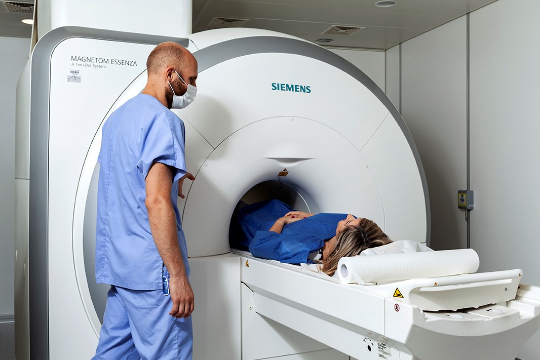
Like all MRIs, breast MRI is a diagnostic technique that uses magnetic fields, which are harmless to health, to provide detailed, three-dimensional images of the anatomy of a specific body part or tissue.
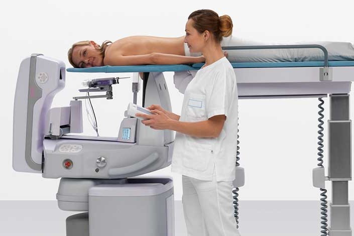
A cylindrical, high magnetic field machine with a bed inside is used to perform it.
In the case of breast MRI, special cup-shaped cavities are provided on the couch where the breasts are positioned.
![]() MRI of the breast with or without contrast medium
MRI of the breast with or without contrast medium
Magnetic resonance imaging of the breast is a 2nd level method that is used according to precise indications defined in international medical and scientific literature.
In some cases, the examination is performed with the use of contrast medium, in others without.
Let’s see specifically.
Breast MRI with contrast medium
MRI with contrast medium is performed in the following cases
- to monitor women with a high familial risk of developing breast cancer, combined with the presence of specific mutations of the BRCA1 and BRCA2 genes;
- to investigate doubts revealed by mammography or ultrasound examination;
- before surgery for local staging of the tumour and to assess the possible existence of additional foci of neoplasia not visible on conventional examinations (mammography and ultrasound);
- in patients who have already been operated on for breast cancer, in whom there is any doubt between a tumour recurrence or surgical scarring resulting from one or more previous operations.
Breast MRI without contrast medium
Without contrast medium, MRI of the breast is used for the evaluation of
- integrity of aesthetic or reconstructive breast implants after oncological surgery;
- possible complications.

How does the examination take place and how long does it last?
After depositing personal items, particularly those sensitive to the magnetic field, such as watches and credit cards, the nurse prepares the venous access by inserting a cannula needle into a vein in the woman’s arm, through which the contrast medium will be administered during the examination.
Then the woman, her chest uncovered, is positioned by the technician on the couch in a prone position with her breasts inside the appropriate cavities.
The examination is painless and lasts about 20-25 minutes.
How to prepare for the examination
In the case of an examination with contrast medium, you must have been fasting for 6 hours and present a creatinine test.
Since no ionising radiation is used, the woman can carry out any activity immediately before the examination and can be in contact with children and frail persons immediately afterwards.
What are the contraindications?
Contraindications are the same as for any MRI examination.
It cannot be performed in the presence of pacemakers or metal prostheses.
Read Also:
Emergency Live Even More…Live: Download The New Free App Of Your Newspaper For IOS And Android
What Is An ECG And When To Do An Electrocardiogram
MRI, Magnetic Resonance Imaging Of The Heart: What Is It And Why Is It Important?