
Melasma, a skin disorder due to hypermelanosis
Melasma consists of (typical and characteristic) zone hyperpigmentation resulting in the formation of visible spots
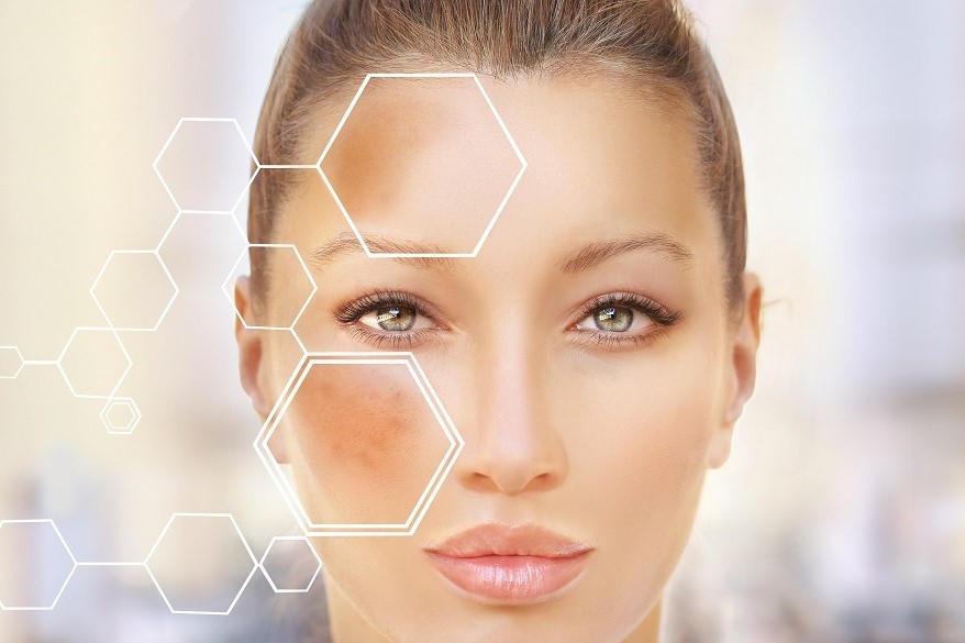
Melasma spots are usually coffee-coloured or tending to grey, irregularly shaped, and are most frequent in the so-called T-zone (forehead, nose and chin) as well as on the cheekbones and upper lip.
They are different from moles because not only are they not raised, but they usually appear very small and then enlarge over time (nearby spots tend to join and form larger ones).
They are asymptomatic and typically worsen after sun exposure precisely because those areas, richer in melanin, tend to tan more than others.
In addition to the face, they can also appear on other areas such as the arms and neck.
The most recent medical research provides very interesting data on the disease.
Those most affected by melasma are female and of childbearing age or during pregnancy.
This is because the dysfunction seems to result directly from the action of particular hormones such as oestrogen and the intake of certain contraceptive drugs.
Genetics and belonging to certain ethnic groups may also have an influence.
Ethnic groups with darker skin tones and greater exposure to sunlight (African ethnicities and island groups) are more affected.
In smaller numbers, men are also affected.
Types of melasma
To date, four different types of melasma have been identified.
In milder cases, the spots are detected, studied and classified by the dermatologist following a dermatoscopic investigation using Wood’s light.
It is different in more pronounced cases, where melanin accumulations are visible even to the naked eye.
Epidermal melasma is the first type and corresponds to the mildest manifestation
It is in its initial stage and the spots are light brown in colour, deposited in the most superficial layer of the skin (the epidermis).
The hair follicles remain intact.
When the dermatoscopic investigation returns the figure of a blue-grey pigmentary network, it means that we are looking at the case history of dermal melasma.
The macrophages containing melanin are deposited in this case in the more superficial dermis (the dermis is the skin layer just below the epidermis and is divided into two parts, one superficial and one deeper).
Mixed melasma, on the other hand, occurs when the dark spots originate in both the epidermis and the superficial dermis.
In melasma of skin phototype V and VI, the lesions are clearly visible in daylight.
In contrast, melasma with telangiectasia is diagnosed when hyperpigmentation also affects the vascular area.
The typical signs of melasma
As already mentioned, melasma is not a dysfunction whose only consequences are severe aesthetic and consequently psychological discomfort.
The typical sign of an accumulation of melanin is the formation of coffee-coloured, dark brown or greyish spots on the skin.
These are usually shades that are noticeable due to the contrast with neighbouring healthy skin.
The spots, which appear in the initial stages as very small, may later coalesce to form larger ones.
They are often irregular, not only in shape but also in internal colouring, as melanin is never evenly deposited in all places.
It is easy to notice the presence of melasma as the spots usually erupt on the face where the preferred areas are the cheekbones, the T-zone (forehead, nose, chin) and the upper lip, usually during pregnancy and, unlike other skin spots, darken with exposure to the sun.
Melasma can also spread to the neck and arms, although more rarely.
Causes
Underlying the appearance of melasma is a localised overproduction of melanin, usually hormonal, genetic or secondary to other systemic diseases.
Staying in the hormonal sphere, it is not uncommon for melasma to be a consequence of high oestrogen production, taking the contraceptive pill or an irregular menstrual cycle.
There is also a special type of melasma that has taken on a name of its own: chloasma (or mask gravidum) is the typical melasma of pregnancy.
The presence of spots with too much melanin can be significantly worsened by excessive exposure to UVA and sunlight.
By sunbathing, the spots tan even more, promoting a colour imbalance with the rest of the skin. In practice, the spots become even darker and more noticeable.
The same can be done by taking photosensitising drugs.
Melasma: how is it diagnosed?
Diagnosing melasma is fairly simple.
An initial visit to one’s general practitioner will, after careful observation, be able to give a quick diagnosis from an objective test and a brief anamnesis.
The dermatologist takes care of the actual specialist test, with the task of observing the change in skin pigmentation and classifying the individual case into the most suitable type of melasma.
Also for melasma, the first anamnesis phase includes the collection of the patient’s clinical information and direct observation of symptoms.
For mild melasma, the objective test consists of observation with Wood’s light, a special technique that highlights skin spots and lesions at different levels of depth, carefully dividing those located in the epidermis from those in the lower layers of the dermis.
Wood’s light is the most useful test if one wants to detect how deep the melanin has reached.
The doctor, after observation with Wood’s light, may decide to undertake a dermoscopy examination of the lesion for a differential diagnosis with melanoma.
This is reserved for the most doubtful or suspicious cases.
Following the diagnosis, it will be up to the doctor to determine the most appropriate methods of intervention, reassuring the patient that melasma is not contagious and cannot evolve into skin cancer.
Treatments for melasma
As we have seen, melasma is often the result of passing periods and hormonal changes in our lives.
This is the reason why many times the disease regresses or resolves itself. Chloasma or melasma gravidarum should disappear spontaneously after childbirth, and the same applies to melasma caused by contraceptive methods, which ends of its own accord after they are discontinued.

To heal hormonal melasma, however, one must act directly by treating the hormonal imbalance that caused it.
In general, there is a wide range of treatments for melasma on the market today, most of which exploit the topical action of certain active ingredients.
Numerous creams, ointments and lotions are available to apply directly on the area of concern, based on arbutin, Vitamin C, glycolic acid and retinoic acid.
These are skin-lightening creams that are only available on prescription.
They can temporarily improve the situation but if the cause is not resolved, the spots are bound to reappear.
The expansion of technologies used in the medical and wellness branch has enabled the arrival of new treatment methods for melasma.
They are based on the use of special types of deep peelings that work by reducing hyperpigmentation.
These techniques counteract the action of melanin and restore a skin tone that is as normal as possible.
The most commonly used reagent is hydroquinone, with exfoliating and melanin-inhibiting action, capable of increasing and speeding up the regeneration of good cells on diseased ones.
However, the first step towards preventing melasma remains conscious sun exposure and the constant use of sunscreens.
Sun protection is not only useful against melasma, but also prevents the onset of skin tumours.
It is also recommended to avoid sunlamps, as direct UV rays on the skin are always harmful in the long run.
Make-up is also increasingly involved in resolving (or rather camouflaging) melasma.
There are many dermatologists and make-up artists who advise patients to rely on skin camouflage techniques and, therefore, on make-up that quickly and directly covers the colour difference given by the spots.
Read Also
Emergency Live Even More…Live: Download The New Free App Of Your Newspaper For IOS And Android
Eczema, Rash, Erythema Or Dermatosis: Let’s Talk About Our Skin
Hyperchromia, Dyschromia, Hypochromia: Skin Color Alterations
Seborrheic Dermatitis: Definition, Causes And Treatment
Dermatosis: Definition, Symptoms, Causes, Diagnosis And Treatment
Allergic Dermatitis: Symptoms, Diagnosis, Treatment
Dermatitis: Causes, Symptoms, Diagnosis, Treatment And Prevention
Skin, What Are The Effects Of Stress
Eczema: Definition, How To Recognise It And Which Treatment To Favour
Dermatitis: The Different Types And How To Distinguish Them
Contact Dermatitis: Patient Treatment
Stress Dermatitis: Causes, Symptoms And Remedies
Infectious Cellulitis: What Is It? Diagnosis And Treatment
Contact Dermatitis: Causes And Symptoms
Skin Diseases: How To Treat Psoriasis?
Eczema Or Cold Dermatitis: Here’s What To Do
Psoriasis, An Ageless Skin Disease
Psoriasis: It Gets Worse In Winter, But It’s Not Just The Cold That’s To Blame
Childhood Psoriasis: What It Is, What The Symptoms Are And How To Treat It
Skin Lesions: Difference Between Macula, Papule, Pustule, Vesicle, Bulla, Phlycten And Wheal
Topical Treatments For Psoriasis: Recommended Over-The-Counter And Prescription Options
Eczema: How To Recognise And Treat It
What Are The Different Types Of Psoriasis?
Phototherapy For The Treatment Of Psoriasis: What It Is And When It Is Needed
Skin Diseases: How To Treat Psoriasis?
Basal Cell Carcinoma, How Can It Be Recognised?
Acariasis, The Skin Disease Caused By Mites
Epiluminescence: What It Is And What It Is Used For
Malignant Tumors Of The Skin: Basal Cell Carcinoma (BCC), Or Basalioma
Chloasma: How Pregnancy Alters Skin Pigmentation
Burn With Boiling Water: What To Do / Not To Do In First Aid And Healing Times
Autoimmune Diseases: Care And Treatment Of Vitiligo
Classification Of Skin Lesions