
Mole mapping: more accuracy with videodermatoscopy
Mole mapping with videodermatoscopy: a more accurate technique that captures microstructural changes in moles and compares them over time
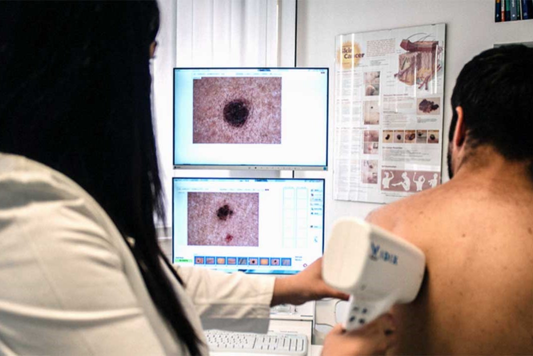
Mole mapping with videodermatoscopy: checking your moles is an essential routine examination for the prevention of skin cancer
By means of a thorough and periodic check-up, it is possible to record any changes in the colour, size and shape of the moles, which may be a premonition of degeneration into melanoma.
This check, which for some subjects must be repeated every year, is important for assessing the nature of the moles and intervening in time.
It can be carried out with the optical dermatoscope, with which the dermatologist analyses the morphology of the enlarged mole, or with the digital videodermatoscope, a new generation machine that allows greater functions and greater precision.
Mole mapping: the advantages of videodermatoscopy
The advantage over the traditional method is that with videodermatoscopy we can:
- acquire the enlarged images on the computer
- archive them;
- compare them with those of previous and subsequent examinations, in order to highlight even minimal changes that could be a sign of tumour development.
With this technology we avoid relying on the doctor’s or patient’s memory, also because microstructural changes in a mole cannot be seen with the naked eye.
The changes that can be seen with the naked eye, and remembered between visits, are only the large ones, with the considerable risk of intervening on a mole late.
High quality images and enlargements, combined with archiving and comparison, allow us to select more accurately who to operate on and who not to.
This does not mean that we resort to multiple operations, quite the contrary.
On the contrary, we avoid unnecessary removals and speed up the removal of seemingly harmless moles.

When to have your moles mapped
My advice is to have a videodermatoscopy at least once in your life.
After the first check-up, two types of patients are identified.
For some, who are more at risk, the images are archived and a check-up is scheduled at 6-12 months if the lesions are particularly irregular.
For the other patients, no images are archived: they are considered low risk and the next visit is scheduled after 2-3 years.
The first dermoscopy can be carried out in adolescence, between 14 and 16 years of age, because one begins to have an idea of the future development of the moles.
If no criticalities appear, the next visit is done after 4-5 years.
Read Also:
What Is A Tumour And How It Forms
Rare Diseases: New Hope For Erdheim-Chester Disease
How To Recognise And Treat Melanoma
Moles: Knowing Them To Recognise Melanoma