
Multiple congenital arthrogryposis: causes, symptoms, diagnosis, treatment
Arthrogryposis (or ‘multiple congenital arthrogryposis’ hence the acronym ‘AMC’; in English ‘arthrogryposis multiplex congenit’ or ‘arthrogryposis’) in medicine refers to a clinical condition characterised by joint stiffness present at birth (hence the name ‘congenital’), in two or more areas of the body (‘multiple’)
The term ‘arthrogryposis’ is derived from the Greek arthron, joint, and grypos, rigid
Cases of arthrogryposis are relatively rare and occur in approximately 1 in 3000 live births.
Amyoplasia, characterised by fatty and fibrous tissue instead of arm muscles, is the most frequent form, occurring in 43% of cases.
The diagnosis and treatment of arthrogryposis can involve various professionals including paediatrician, orthopaedist, neurologist, child neuropsychiatrist, physiatrist and physiotherapist.
Causes and risk factors of arthrogryposis
The specific causes of arthrogryposis have not yet been fully elucidated, however it seems clear to researchers that anything that inhibits normal fetal joint movement (i.e. before birth) can cause the proliferation of collagen fibres and the replacement of muscles with fibrous tissue leading to joint contractures.
Put simply: any factor that restricts fetal movement can cause congenital contractures.
Among the causes that could determine or favour arthrogryposis, we include neurological, myopathic and intrauterine pathologies.
Myasthenia gravis in the mother also leads to arthrogryposis in rare cases.
The main cause in humans according to some is foetal akinesia.
Arthrogryposis could be caused by environmental (extrinsic), intrinsic and genetic factors.
Extrinsic factors
Arthrogryposis malformations may be secondary to environmental factors such as:
- reduced intrauterine movement
- oligohydramnios (low volume or abnormal distribution of intrauterine fluid);
- defects in fetal blood supply;
- hyperthermia;
- immobilisation of limbs;
- viral infections;
- disorders of muscle and connective tissue development such as muscular dystrophy, various types of myopathy and mitochondrial disorders.
Seventy per cent of cases of the most severe forms of arthrogryposis appear to be related to neurological abnormalities.
Intrinsic Factors
Arthrogryposis could also be caused by intrinsic factors including developmental disorders of molecular, muscular and connective tissue or neurological abnormalities.
Genetic causes of arthrogryposis
There appear to be more than 35 specific genetic disorders associated with arthrogryposis.
Most of these mutations are missense, meaning that the mutation causes a different amino acid.
Other mutations that could cause arthrogryposis are single gene defects (X-linked recessive, autosomal recessive and autosomal dominant), mitochondrial defects and chromosomal disorders (e.g. trisomy 18); this is mainly found in distal arthrogryposis.
Mutations in at least five genes (TNN12, TNNT3, TPM2, MYH3 and MYH8) may cause distal arthrogryposis.
Arthrogryposis is divided into two groups:
- amyoplasia: the most common form of arthrogryposis, characterised by rather symmetrical and typical limb involvement, severe joint contractures, muscle weakness, normal intelligence, and frequently an angioma in the face
- distal and syndromic arthrogryposis: a group of disorders characterised by autosomal dominant transmission and greater involvement of the hands and feet; Freeman-Sheldon syndrome or whistler syndrome and Beals syndrome or distal arthrogryposis type 9 belong to the latter group.
Symptoms and signs
Babies born with one or more joint contractures have abnormal fibrosis of the muscle tissue that causes muscle shortening and therefore are unable to perform active extension and flexion in the affected joint(s).
Often every joint in a patient with arthrogryposis is affected:
- in 84% of cases all limbs are involved;
- in 11% only the legs are involved;
- in 4% only the arms are involved.
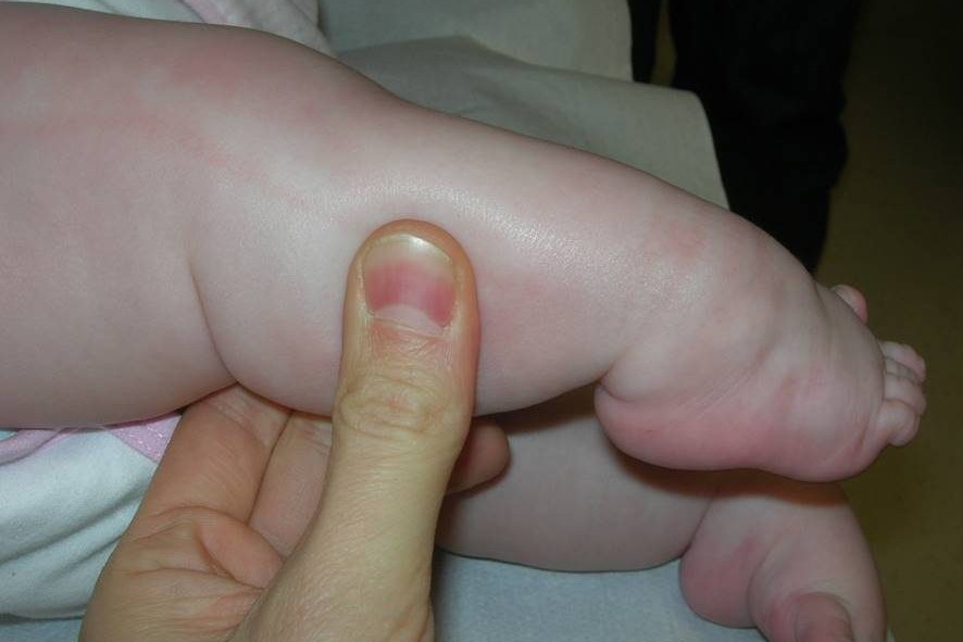
Each joint of the body, when affected, shows typical signs and symptoms: e.g. shoulder (internal rotation); wrist (volar and ulnar); hand (fingers in fixed flexion and thumb in the palm); hip (flexed, abducted and externally rotated, often dislocated); elbow (extension and pronation) and foot (club foot).
The range of motion may differ between the various joints involved, due to the different deviations.
Some types of arthrogryposis such as amyoplasia have symmetrical involvement of the limb joints. Sensitivity is generally normal.

Joint contractures can cause delayed gait development in the first five years of life, but the severity of the contractures does not necessarily predict the eventual ability to walk or inability to walk: each case can have a completely different course depending on the severity of the situation.
The intelligence of children with amyoplasia is normal.
Some syndromes, such as Freeman-Sheldon and Gordon syndrome, have craniofacial involvement.
The amyoplasia form is sometimes accompanied by a midline facial haemangioma.
Arthrogryposis is often accompanied by other syndromes or diseases, so the other symptoms and signs present at the same time can be extremely varied, depending on the pathology from which the child suffers.
Frequently, the child with arthrogryposis also suffers from:
- pulmonary hypoplasia with dyspnoea and respiratory insufficiency;
- cryptorchidism;
- congenital heart defects with frequent alterations detectable on the electrocardiogram;
- tracheoesophageal fistulas;
- inguinal hernias;
- cleft palate;
- hypotonia of the infant or floppy infant syndrome;
- eye abnormalities.
Arthrogryposis can be detected by objective examination and confirmed by ultrasound, MRI and muscle biopsy
Research into prenatal diagnosis has shown that a diagnosis can be made prenatally in approximately 50% of foetuses presenting with arthrogryposis: the latter could be detected during a normal prenatal ultrasound scan, which shows a lack of mobility and/or an abnormal position of the foetus.
4D ultrasound may be used.
Therapy
The treatment of children with arthrogryposis must be organised by a multidisciplinary team of experts and must take into account different aspects (motor and walking aspects, communication skills, activities of daily living, independence.
Different treatment tools exist, including:
- physiotherapy (stretching, joint mobilisation…) to be started immediately after birth;
- occupational therapy;
- orthopaedic braces;
- orthopaedic footwear;
- corrective surgery such as dorsal carpal wedge oseotomy or index rotation flap.
The primary long-term goals of these treatments are to increase joint mobility, muscle strength and the development of adaptive use patterns that allow for walking and independence with activities of daily living.
Since arthrogryposis comprises many different types, treatment varies from patient to patient.
Since arthrogryposis comprises many different types and can have different severity, the prognosis can be very variable
It must be said that the joint contractures present will not worsen over time any more than they did at birth, however there is no therapy that can cure arthrogryposis completely.
In general, arthrogryposis is not a progressive condition, so with proper medical rehabilitation treatment, the situation can certainly improve: most children have significant improvements in their range of motion and ability to move their limbs, allowing them to carry out activities of daily living and live a relatively normal life.
In more severe cases, surgery can improve mobility and joint function, allowing the subject to be independent.
In some cases, normal walking is made extremely difficult or completely impossible.
Positive prognostic factors for independent walking (without a wheelchair) are hip flexion contractures of less than 20° and knee flexion contractures of less than 15° in the absence of severe scoliosis.
Read Also:
Emergency Live Even More…Live: Download The New Free App Of Your Newspaper For IOS And Android
How To Survive The Witch’s Stroke: Discovering Acute Low Back Pain
Lumbago: What It Is And How To Treat It
Back Pain: The Importance Of Postural Rehabilitation
Epiphysiolysis: ‘Train Paediatricians To Avoid Late Diagnoses’
Idiopathic Scoliosis: What It Is And How To Treat It
Diagnosis And Treatment For Adult Scoliosis
Is Correcting Scoliosis Possible? Early Diagnosis Makes All The Difference