
Oncology, an overview of orbital tumours
Orbital tumours constitute a large group of primary lesions (i.e., originating from the orbit of the eye), secondary lesions (originating from regions contiguous to the orbit: eyeball, ocular adnexa, paranasal sinuses, nasopharynx) and metastatic lesions (originating from areas of the body distant from the orbit)
They are divided into neurogenic, mesenchymal, vascular, epithelial lacrimal gland tumours, lymphoproliferative lesions, secondary tumours and metastases.
What are orbital tumours?
Neurigenic tumours (optic nerve glioma and optic nerve meningioma) involve the peripheral nerves and may originate from different cellular components (Schwann cells, axons, fibroblasts and nerve sheaths).
Mesenchymal tumours (such as rhabdomyosarcoma, leiomyoma, lipoma, liposarcoma, fibromas, fibrosarcomas, solitary fibrous tumour and fibrohistiocytoma) originate from mesenchymal tissue, are rare and vary from slowly progressing benign lesions to severe inflammatory vascular lesions and destructive malignancies.
Lymphoproliferative lesions of the orbit range from reactive lymphoid hyperplasia to malignant lymphoma and in most cases are non-Hodgkin lymphomas arising from B lymphocytes.
Finally, secondary tumours originate from the eyeball and ocular adnexa and may spread, for example, through the optic nerve (such as choroid melanomas), while neoplasms that most frequently metastasise in the orbit originate from the breast, lung, prostate, melanoma, colon and kidney.
What are the symptoms of orbital tumours?
Each type of tumour is characterised by symptoms ranging from protrusion in the eyeball beyond the orbit to palpebral ptosis.
Vision may diminish and, in some cases, double.
The correct diagnosis of an orbital tumour may require:
Comprehensive eye examination
– Radiological imaging to localise and quantify the tumour mass
– Multidisciplinary consultation between oncology specialists and neurosurgeons in the case of glioma and meningioma and between haematologists and anatomopathologists in the case of lymphoproliferative lesions, secondary tumours and metastases
– Cv
– Orthoptic examination
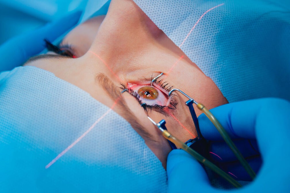
Treatments
If vision is good the optic nerve glioma only needs to be carefully monitored with an eye examination and MRI of the brain or orbits.
In more serious cases, surgical removal is necessary.
The same applies to meningioma, which requires surgery only if the tumour reaches the optic canal.

Treatment of lymphoproliferative lesions involves radiotherapy for localised lesions and chemotherapy for disseminated disease.
Unfortunately, there are no preventive measures.
Read Also:
Emergency Live Even More…Live: Download The New Free App Of Your Newspaper For IOS And Android
Corneal Keratoconus, Corneal Cross-Linking UVA Treatment
Myopia: What It Is And How To Treat It
Presbyopia: What Are The Symptoms And How To Correct It
Nearsightedness: What It Myopia And How To Correct It
Blepharoptosis: Getting To Know Eyelid Drooping
Lazy Eye: How To Recognise And Treat Amblyopia?
What Is Presbyopia And When Does It Occur?
Presbyopia: An Age-Related Visual Disorder
Blepharoptosis: Getting To Know Eyelid Drooping
Rare Diseases: Von Hippel-Lindau Syndrome
Rare Diseases: Septo-Optic Dysplasia
Diseases Of The Cornea: Keratitis
Inflammations Of The Eye: Uveitis