
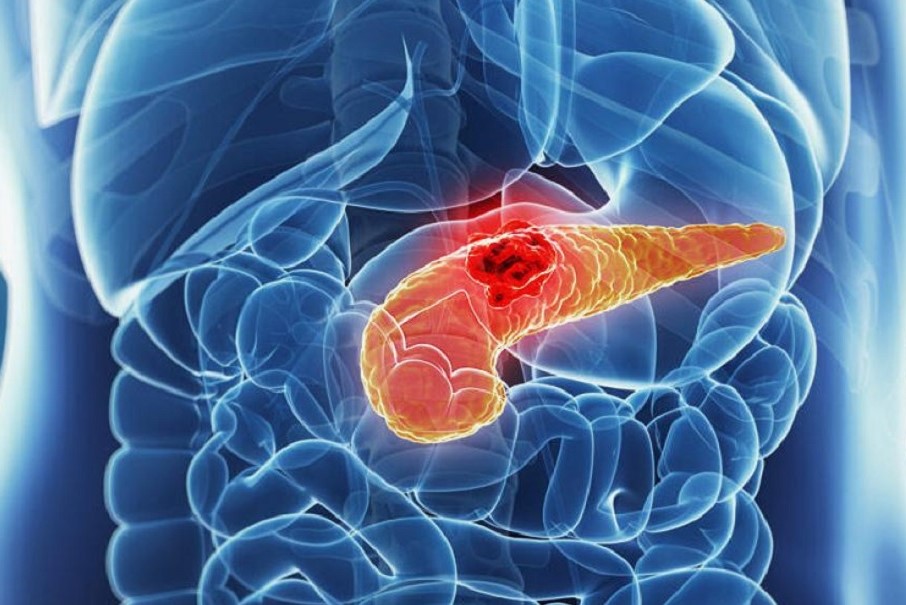
Pancreas: prevention and treatment of pancreatic cancer
There are several other types of pancreatic cancer, but adenocarcinoma is the most common pancreatic neoplasm
It is a disease that originates in the ducts that carry digestive enzymes produced by the pancreas, the causes of which are still unknown.
It forms as a result of the accumulation of pancreatic cells that have undergone a genetic alteration that causes them to multiply uncontrollably, without ever dying, and that grow and spread rapidly.
This is why it is a difficult neoplasm to diagnose at an early stage.
Types of pancreatic exocrine tumours
About 80% of pancreatic exocrine tumours are adenocarcinomas of the ductal epithelium, and only 2% are benign.
There are some very rare exocrine tumours such as giant cell carcinoma, adenosquamous carcinoma, microglandular adenocarcinoma, mucinous carcinoma, cystadenocarcinoma, papillary cystic carcinoma, cystadenocarcinoma and acinar cell cystadenocarcinoma, and finally primary pancreatic lymphoma (tumour starting in connective tissue).
Cystic tumours account for less than 5% of all pancreatic tumours (early mucinous cystadenoma and cystadenocarcinoma), while intraductal and mucinous tumours (both benign and malignant) generally occur as a cystic dilatation of the pancreatic ductal system.
Endocrine tumours, which are initiated by pancreatic duct cells, may be functionally inactive, benign or malignant functioning, and include insulinomas, glucagonomas and gastrinomas.
Approximately 40% of pancreatic endocrine tumours are non-functional and of these up to 90% are malignant
There are several syndromes that include neoplasms associated with pancreatic tumours.
Of these, the best known is multiple endocrine neoplasia (MEN) type 1 (parathyroid hyperplasia, endocrine pancreatic tumours and pituitary tumours), and gastrinomas and insulinomas are most frequently represented here.
These variations in tumour function influence diagnosis and treatment strategies.
Clinical strategy and treatment for pancreatic cancer
When the diagnosis of pancreatic cancer is made, it is necessary to assess whether it is amenable to radical surgical resection.
A pancreatic tumour in general terms is defined as resectable when it can be removed without leaving neoplastic residue (R0), unresectable when it cannot be completely resected (R1-2) or borderline when radicality is doubtful preoperatively and one has to rely on neoadjuvant therapies or directly on surgical evaluation during surgery.
Unfortunately, only about 20% of patients with pancreatic cancer at the time of diagnosis are radically resectable.
Therefore, it is crucial to define the tumour staging according to the TNM system (AJCC) as precisely as possible to avoid unnecessary interventions.
The AJCC staging of a pancreatic cancer is as follows:
Tumour (T)
TX – Primary tumour cannot be assessed
T0 – No evidence of primary tumour
Tis – Carcinoma in situ
T1 – Tumour limited to the pancreas, 2 cm or smaller in maximum size
T2 – Tumour limited to the pancreas, larger than 2 cm in greatest dimension
T3 – Tumour extends beyond the pancreas (e.g. duodenum, bile duct, portal or superior mesenteric vein) but does not involve the celiac axis or superior mesenteric artery
T4 – Tumour involves the celiac axis or superior mesenteric arteries
Regional lymph nodes (N)
NX – Regional lymph nodes cannot be assessed
N0 – No regional lymph node metastases
N1 – Regional lymph node metastases
Distant metastases (M)
MX – Distant metastases cannot be assessed
M0 – No distant metastases
M1 – Distant metastases
The stage grouping for pancreatic cancer is as follows:
Stage 0 – Tis, N0, M0
Stage IA – T1, N0, M0
Stage IB – T2, N0, M0
Stage IIA – T3, N0, M0
Stage IIB – T1-3, N1, M0
Stage III – T4, Any N, M0
Stage IV – Any T, Any N, M1
At initial presentation, only 20% of patients have stage I disease, 40% have locally advanced disease and 40% have metastatic disease to lymph nodes or distant sites.
Generally speaking, T1 and T2 stages are confined to the pancreatic parenchyma, whereas T3 lesions invade local structures such as the duodenum, bile duct and/or major peripancreatic veins and T4 lesions invade surrounding organs (e.g. stomach, colon, liver) or major arteries such as the superior mesenteric or celiac arteries.
Pre-operative staging laparoscopy
Some centres advocate performing a staging laparoscopy before proceeding to laparatomy.
The purpose of laparoscopic staging is to avoid subjecting patients with liver or peritoneal metastases not visible with common radiological methods to unnecessary surgery.
Such an investigation is however appropriate when these situations are present:
- CA 19-9 level > 150 U / mL
- Low volume ascites
- Tumours in the body of the pancreas
- Borderline resectable tumours
- Tumour size> 3 cm
- Common bile duct lymphadenopathy
- Therapy and treatment of pancreatic cancer in operable forms
There is a reasonable consensus in the literature that surgery, when radically possible, is the main treatment modality for pancreatic cancer.
However, there is an important role for chemotherapy and/or radiotherapy in an adjuvant or neoadjuvant setting and in the treatment of patients with unresectable disease.
Generally, extrapancreatic disease precludes curative resection and surgical treatment can be palliative at best.
Generally, vascular infiltration is considered a contraindication to pancreatic resection, although nowadays invasion of the mesenteric or superior portal vein is no longer an absolute contraindication, in fact the latter can be removed and reconstructed (moreover, in many cases the vein is only compressed and not infiltrated) using the internal jugular vein, great saphenous vein or splenic vein.
The assessment of the infiltration of the superior mesenteric, celiac and hepatic arteries, on the other hand, is different, as they indisputably represent an absolute contraindication to radical resection.
In this context, the surgical approach must be adapted to the tumour site and therefore duodenocephalopancreasectomy (DCP), with or without pylorus sparing, total pancreasectomy (PT) and distal pancreasectomy (PD) will be considered.
These procedures, which require surgeons experienced in this type of surgery to perform, have an incidence of complications and preoperative risks that must be known and discussed with the patient before proceeding with the operation.
The pancreatic cancer guidelines of the European Society of Medical Oncology (ESMO) indicate that complete surgical resection is the only potentially curative treatment available; however, 5-year overall survival is only 10-20%, and long-term survival in patients with lymph node spread disease is extremely poor.
ESMO recommendations include the following
Optimal symptomatic treatment plays a key role in the management of metastatic disease; patients may require drainage or bypass surgery for obstructive jaundice or gastric obstruction.
The role of chemotherapy is limited. Gemcitabine has been associated with a small survival benefit compared to bolus 5-fluorouracil.
In patients with locally advanced unresectable pancreatic cancer, local ablation has been explored as a therapeutic option. A systematic review concluded that the following strategies appear to be feasible and safe:
- Radiofrequency ablation (RFA)
- Irreversible electroporation
- Stereotactic body radiotherapy (SBRT)
- High intensity focused ultrasound (HIFU)
- Iodine-125
- Iodine-125-cryosurgery
- Photodynamic therapy
- Microwave ablation
- Many of these ablative techniques have been shown to provide pain relief and improved survival.
For example, medial survival of up to 25.6 months has been reported with RFA and 24.0 months with SBRT. Promising results on quality of life have been reported for SBRT.
The other procedures have shown promising results in some facilities, but still have low uptake.
Chemotherapy for pancreatic cancer
There are currently no treatment protocols that give guarantees of success in this area.
It is believed that in patients with metastatic disease, the combination of Gemcitabine and Erlotinib can provide significantly higher median survival and 1-year survival than the use of Gemcitabine alone.
Some studies indicate that the Gemcitabine-Capecitabine combination is one of the standard first-line options in locally advanced and metastatic pancreatic cancer, while other studies have shown that the FOLFIRINOX combination (leucovorin plus 5-lfuourouracil [LV5-FU] plus oxaliplatin plus irinotecan) is able to promote a median survival of 11.1 months compared to 6.8 months in the Gemcitabine alone group.
There are currently no uniformly accepted and agreed protocols.
Adjuvant therapy
Several studies have suggested the possibility that chemotherapy, with or without radiotherapy, may significantly improve median survival after surgical resection of operable disease.
These studies are not definitive and are not widely accepted to justify chemo-radiotherapy treatment for adjuvant therapy.
Neoadjuvant therapy
The use of chemotherapy and/or radiotherapy in the neoadjuvant setting is also still a source of controversy.
The rationale for the use of neoadjuvant therapy includes the assertions that:

- pancreatic cancer is a systemic disease and must be treated systematically from the outset;
- patients are able to tolerate the toxic effects of chemotherapy more readily before undergoing major pancreatic resection afterwards;
- the tumour may shrink in size with neoadjuvant therapy and resection may be less complex, leading to better overall survival.
The problem is that there is still no definitive agreement on which treatment protocols to use in this clinical setting.
Duodenocephalopancreasectomy (Whipple’s DCP-procedure)
This operation is performed in patients presenting with a neoplasm of the head of the pancreas, Vater’s papilla or terminal choledoch or duodenum.
The operation traditionally involves the removal of the pancreatic head, duodenum, gallbladder and antrum of the stomach, with surgical drainage of the distal pancreatic duct and the biliary system, usually performed by anastomosis with a digiunal loop (biliary-digestive anastomosis).
DCP has been shown to have an overall mortality rate of 6.6% and a morbidity rate of 25%.
The most serious complications are anastomotic fistulas, delayed gastric outlet and digestive haemorrhage.
In patients with jaundice, many authors propose the use of biliary drainage (endoscopic or transhepatic) placed preoperatively.
However, to be effective, biliary drainage must be maintained for an adequate time to normalise or nearly normalise the bilirubin level (approximately 20 days).
However, this solution, in addition to delaying the operation, predisposes to the risk of infection of the biliary tract, which in turn is associated with an increased risk of postoperative infectious complications and wound infection, and for these reasons does not find convinced supporters in the majority of authors.
Whipple’s standard operation can be modified by avoiding resection of the gastric antrum by preserving the pylorus.
This modification has been proposed to improve the nutritional status of the patient (reservoir function of the stomach), but may be burdened by an increased risk of slowing gastric emptying (removal of the duodenum-gastric innervation).
From an oncological point of view, there are no differences between the two procedures.
Pancreatic cancer, the guidelines of the European Society of Medical Oncology give the following recommendations
- surgical resection with radical intent is the only potentially curative treatment available. However, 5-year overall survival is only 10-20%; long-term survival in tumours with positive lymph nodes is extremely poor.
- Optimal symptomatic treatment plays a key role in the management of metastatic disease; these patients may require drainage or bypass surgery for obstructive jaundice or gastric outlet obstruction.
- The role of chemotherapy is limited; Gemcitabine alone or in combination with other drugs has been associated with a small survival benefit.
Distal pancreatectomy (PD)
This procedure has a lower mortality rate than Whipple’s standard procedure, at 3.5%, but its use in curative resection remains limited.
PD is effective and technically less complex than DCP in distally located tumours.
Unfortunately, masses located in this area are diagnosed much later and therefore are usually easily inoperable due to vascular thrombosis or gastric or intestinal infiltration.
The procedure involves isolation of the distal portion of the pancreas containing the tumour, followed by resection of that segment with or without the spleen, with suturing of the distal pancreatic duct.
The main complications for PD are pancreatic fistula, haemorrhage and an abscess.
It is essential to place adequate drains of the area involved in the surgical act.
Total pancreasectomy (PT)
Although this procedure is the least commonly performed, it may still be a valuable tool in the surgical treatment of pancreatic cancer, particularly in cases where the tumour involves the neck of the pancreas or there is a suspicion that the tumour has metastasised along the duct of Wirsung.
The mortality rate is around 8% and the morbidity is lower (in fact, there is no risk of pancreatic fistulas or anastomotic dehiscence), but it results in a definitive postoperative total diabetes state that is not always easy to treat.
Palliative therapy for pancreatic cancer
Pain
In patients who are not amenable to surgical resection, it is necessary to offer therapy to prevent and treat the most significant and disabling symptoms of the disease.
Pain relief is crucial in this context.
The use of narcotic analgesics should be proposed early and at appropriate dosages in combination with tricyclic antidepressants or antiemetics to enhance their analgesic effects.
In patients in whom narcotics are insufficient, other approaches should be considered, such as coeliac ganglion neurolysis, which can provide significant long-term pain relief.
This can be performed transthoracically or transabdominally using interventional radiology or anaesthesiology, trans-gastrically using a fine-needle injection under ultrasound or CT guidance.
Or intra-operatively during evaluation of the patient’s resection potential.
Radiotherapy for pancreatic cancer can relieve pain, but it does not affect patient survival.
Some patients may experience pain due to obstruction of the pancreatic or bile ducts, especially if the pain worsens significantly after eating.
These patients may benefit from endoscopic decompression with pancreatic drainage placed endoscopically retrograde.
Jaundice
Obstructive jaundice is a serious complication for the patient with pancreatic cancer of the head, as it results in intractable itching, which causes severe scratching lesions.
The resolution of this symptom can be achieved by the placement of an internal (transtumoral) biliary drain introduced endoscopically retrograde, internal external (with an external and an internal transtumoral branch) or external (when the tumour is not transitable) by means of a transcutaneous radiological procedure.
Metal expansion prostheses are more expensive and permanent, have a longer patency period and are preferable in patients with an estimated life expectancy of more than 3 months.
Plastic ones are much cheaper and usually have to be replaced every 3-4 months and are preferably used in patients with a shorter life expectancy.
In patients in good general condition, these procedures can also be performed surgically, by means of a bilio-digestive anastomosis (to bypass the pancreatic head neoplasm), a gastro-digiunal anastomosis to obviate duodenal obstruction and ensure nutritional continuity, and celiac neurolysis to prevent pancreatic pain.
Diet for patients with pancreatic cancer
As with most patients with advanced cancer, pancreatic cancer patients are often anorexic.
Pharmacological appetite stimulation is usually unsuccessful, but can be tried.
Patients may present with some degree of malabsorption secondary to exocrine pancreatic insufficiency caused by the cancer obstructing the pancreatic duct.
Patients with malabsorption diarrhoea and weight loss may benefit from pancreatic enzyme supplementation.
Their diarrhoea can also be improved by avoiding high-fat or high-protein diets.
Prevention of pancreatic cancer
Epidemiological data show that smoking is the cause of about 30% of pancreatic tumours.
Specific tobacco carcinogens are methylnitrosamines, nitrosonornicotines, polycyclic aromatic hydrocarbons and aromatic amines.
Various studies have shown that cigarette smokers develop pancreatic cancer 3.3 to 9.5 years earlier than non-smokers.
Cigarette smokers have a 70% higher risk of pancreatic neoplasms than non-smokers.
Cigarettes with a smoke filter do not reduce the risk of cancer.
Not smoking or quitting smoking for good should be considered as a significant cancer prevention factor.
Patients with alcohol-related chronic pancreatitis, if they are long-term smokers at the same time, also have an increased risk of pancreatic and oesophageal cancer.
Surgical removal of pancreatic cancer may also have a preventive aspect in patients with chronic alcoholic pancreatitis.
Since approximately 10% of all pancreatic tumours develop on the basis of chronic hereditary pancreatitis and a familial genetic predisposition, regular screening examinations (EUS, CT scan, MRI) every two to four years are under discussion for these risk groups from the age of 40.
No evidence has yet been provided for the effectiveness of screening examinations.
However, experience with the use of prophylactic resection in high-risk pancreatic cancer is so far only available for a small number of patients.
Patients with cystic neoplasms of the pancreas frequently develop pancreatic cancer in the long term
With IPMN tumours (Intraductal Papillary Mucinous Pancreatic Neoplasms), malignant transformation into ductal pancreatic cancer is observed in approximately 60% -70% of patients.
In IPMN neoplasia, the carcinoma is predominantly located in the head of the pancreas.
Mucinous cystic neoplasms show malignant transformation in about 20% of cases.
Although the increased knowledge of sequential genetic mutations in cystic neoplasia of the pancreas does not yet allow reliable risk prediction, experience has shown that surgical removal is indicated for cystic neoplasms of more than 2 or 3 cm, especially when the diagnosis is known (IPMN, MCN, and serous cystic adenoma).
In many of these diseases, complete removal of cystic neoplasms is a cancer prevention strategy and is now performed without surgical mortality in specialised centres.
The complete removal of a cystic tumour (IPMN, MCN) offers patients a cure for the cystic neoplasm and relieves them of the fear of developing pancreatic cancer.
Read Also:
Emergency Live Even More…Live: Download The New Free App Of Your Newspaper For IOS And Android
Gestational Diabetes, What It Is And How To Deal With It
Pancreatic Cancer, A New Pharmacological Approach To Reduce Its Progression
What Is Pancreatitis And What Are The Symptoms?
Kidney Stones: What They Are, How To Treat Them
Acute Pancreatitis: Causes, Symptoms, Diagnosis And Treatment