
Diabetic retinopathy: the importance of screening
Diabetic retinopathy is the 4th leading cause of blindness worldwide and the leading cause of blindness in the elderly in industrialized countries
It is therefore a relevant social problem, so much so that the World Health Organization has included it in the priority list of preventable diseases, giving specific guidelines for its screening.
In fact, the importance of screening is very high: it is estimated that diabetic patients who do not undergo regular screening have a 4-fold increased risk of developing severe retinopathy.
The main cause of diabetic retinopathy: diabetes
As the name implies, the cause of diabetic retinopathy is diabetes, a condition that affects more than 415 million worldwide, a number estimated to rise to 642 million by 2040.
There are 2 forms of diabetes:
- type 1 (insulin-dependent, IDDM), which is more typical of younger age;
- type 2 (non-insulin-dependent, NIDDM), which normally appears in adulthood.
In each case, it is a chronic and slowly progressive disease that induces complications in several target organs:
- mainly kidneys (nephropathy, chronic renal failure with the need to undergo dialysis);
- heart (the risk of cardiovascular disease is 2 to 4 times higher in people with diabetes than in the rest of the population and is responsible for more than half of all diabetes-related deaths);
- central nervous system (peripheral neuropathy, atrial fibrillation);
- eye: at the ocular level, diabetes leads to consequences especially at the damage of the retina. One third of all diabetics have retinopathy, and the prevalence of diabetic forms of retinopathy that impair visual acuity is 7.9 percent.
Risk factors for the progression of diabetic retinopathy
The main risk factors for the development and progression of diabetic retinopathy are:
- advanced age;
- duration of diabetes (before 5 years of disease, diabetic retinopathy has a negligible prevalence, to reach over 60% of patients after 20 years of disease in type 2 diabetes, reaching peaks of 97% in type 1 diabetes);
- poor glycemic compensation;
- concomitant hypertension;
- particular life stages such as pregnancy and puberty as they are subject to hormonal changes related to increased insulinemic resistance.
Among these risk factors, glycemic compensation is the most important: maintaining good glycemic control (glycated hemoglobin less than 7) in fact reduces the risk of development and progression of diabetic retinopathy.
What is diabetic retinopathy
In terms of the mechanism of action, diabetic retinopathy is a neurovascular disease: it affects the neuronal and endothelial cells of the retina.
Damage to these cells leads to:
- closure of capillary vessels with retinal ischemia that begins in the peripheral part of the retina and extends toward the center (macula);
- accumulation of fluid in the central region of the retina itself (macular edema).
Complications
Progressive ischemia leads to the formation of neovases, which can bleed causing intraocular hemorrhage (called hemovitreo), resulting in acute vision loss.
Sometimes this event resolves with spontaneous reabsorption of the blood; other times surgical removal of the vitreal hemorrhage by vitrectomy is necessary.
Over time, untreated neovases become fibrotic and can lead to retinal detachment, a serious complication that results in abrupt visual decline and requires complex emergency surgery, often followed by failure to recover or partial recovery of vision.
These vessels can also grow on the surface of the iris (the colored part of the eye) and lead to pictures of iris rubeosis (presence of capillaries on the iris) and so-called neovascular glaucoma, a form of glaucoma characterized by a major increase in intraocular pressure with irreversible damage to the optic nerve followed by blindness and pain.
This is a complication that is unlikely to be cured by medical and surgical therapies.
It is evident from what has been said so far that diabetic retinopathy is a very insidious disease
It becomes symptomatic only when the pathology reaches the region of the macula, or when the severe complications that follow ischemia and the proliferating phase of the disease occur so at an already advanced stage.
This is also why a careful and early screening program is essential.
We have many weapons to make early diagnosis of diabetic retinopathy and to monitor it as best we can.
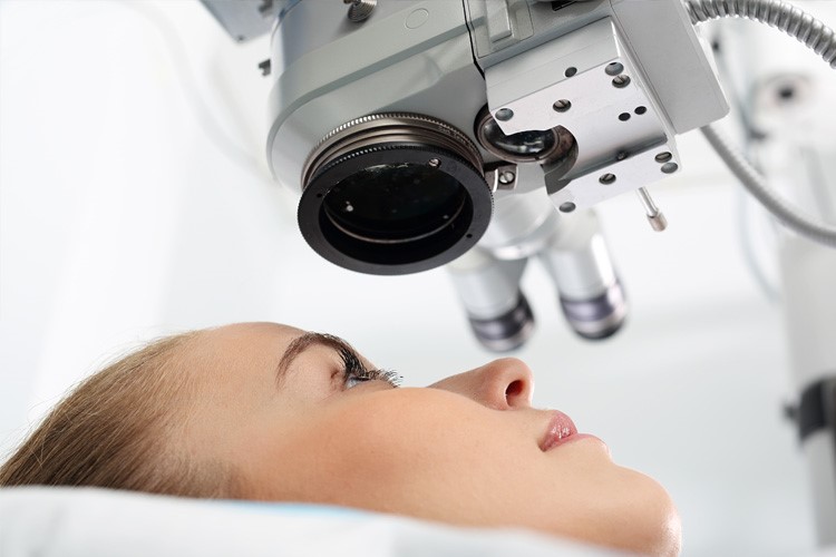
Screening, in particular, is based on ocular fundus analysis.
The first evaluation should be performed:
- after 5 years of diagnosis of type 1 diabetes;
- at the immediate diagnosis of type 2 diabetes.
The interval between follow-up visits is decided by the specialist based on the presence or absence of diabetic retinopathy and its severity.
New instrumentation for diagnosis
The diagnostics of retinal diseases, including that of diabetic retinopathy, has greatly advanced in recent years: today there are instruments that allow in a single visit an accurate assessment of all aspects of this disease.
The diagnostic pathway involves the performance of:
- fluorangiography;
- OCT;
- autofluorescence fundus examination;
- angio-OCT.
Each of these examinations gives us a piece of the ‘puzzle’ for proper final evaluation.
With fluorangiography we assess the presence and extent of retinal ischemia and the presence of neovases
OCT, on the other hand, is the exam that allows us to assess increased macular thickness due to fluid accumulation (macular edema), as well as the presence of tractional epiretinal membranes (‘nonelastic,’ fibrotic tissue that is able to exert traction on the macula leading to edema formation or puncturing it in the center with severe damage on central vision) on the retina that may require surgical intervention.

Finally, autofluorescence allows the study of macular edema, while angio-OCT studies macular ischemia, edema, and shows changes even in the subclinical phase, that is, before symptoms occur.
These examinations also make it possible to check the response to possible therapies and monitor the progress of the disease.
How diabetic retinopathy is treated
The first step of therapy is careful monitoring of the underlying pathology, namely diabetes, motivating and informing the patient about the importance of maintaining good blood glucose levels.
The second step is a good preventive campaign that in the overall evaluation of the diabetic patient makes use of fundus examination and new generation imaging with macula study (OCT, angio- OCT and FAF).
In case of macular edema
When the disease has caused visual decline because it has affected the macula (macular edema), we have the less invasive but effective techniques of ‘grid’ or direct laser photocoagulation used for a long time in treating the edema itself:
- the so-called ‘subthreshold’ laser, a special yellow light laser that allows the treatment of diabetic macular edema through the use of low energies (subthreshold micropulsed) reserved for initial edema with little increase in foveal thickness;
- intravitreal drug injections directly into the eye based on anti-VEGF or steroids when edema is more pronounced.
Help in the early stages of macular edema is provided by supplements containing turmeric and similar substances.
These new therapies often allow the recovery of decent central vision with improvement in the quality of life of our patients.
Diabetic retinopathy, in case of retinal ischemia and neovase formation
When the disease affects the mid-peripheral part of the retina with ischemia and neovases, the treatment of choice is sector laser photocoagulation (for localized ischemia in one area of the retina) or panretinal (affecting all sectors when the damage is more extensive).
This treatment aims to slow down the disease and prevent the onset of serious complications.
In conclusion, today we can try to control the devastating effects of diabetic retinopathy through:
- good metabolic compensation;
- an early diagnosis with a codified screening program;
- constant follow-up using the various tools we have at our disposal;
- when necessary, targeted therapy aimed at preventing progression of the disease to its many complications.
Raising awareness among diabetic patients is very important because we need their help first and foremost to achieve the best results.
Read Also:
Emergency Live Even More…Live: Download The New Free App Of Your Newspaper For IOS And Android
Diabetic Retinopathy: Prevention And Controls To Avoid Complications
Diagnosis Of Diabetes: Why It Often Arrives Late
Diabetic Microangiopathy: What It Is And How To Treat It
Diabetes: Doing Sport Helps Blood Glucose Control
Type 2 Diabetes: New Drugs For A Personalised Treatment Approach
The Diabetic Diet: 3 False Myths To Dispel
Paediatrics, Diabetic Ketoacidosis: A Recent PECARN Study Sheds New Light On The Condition
Orthopaedics: What Is Hammer Toe?
Hollow Foot: What It Is And How To Recognise It
Occupational (And Non-Occupational) Diseases: Shock Waves For The Treatment Of Plantar Fasciitis
Flat Feet In Children: How To Recognise Them And What To Do About It
Swollen Feet, A Trivial Symptom? No, And Here’s What Serious Diseases They May Be Associated With
Varicose Veins: What Are Elastic Compression Stockings For?
Diabetes Mellitus: Symptoms, Causes And Significance Of The Diabetic Foot
Diabetic Foot: Symptoms, Treatment And Prevention
Type 1 And Type 2 Diabetes: What Are The Differences?
Diabetes And Cardiovascular Risk: What Are The Main Complications
Diabetes: Causes, Symptoms And Complications