
Eye diseases: retinitis pigmentosa
Retinitis pigmentosa is a disease that affects the retina. The retina is the structure responsible for receiving light stimuli and converting them into electrical impulses that are then sent to the primary visual cortex (located in the occipital lobe) which has the task of processing and integrating them to reconstruct the image
The retina performs its functions thanks to a complex architecture characterised by the presence of microscopic structures called photoreceptors.
These can be divided into cones and rods and their involution, by mechanisms of apoptosis (programmed cell death), leads to the development of retinitis pigmentosa.
The retina progressively loses its photosensitive capacity.
Retinitis pigmentosa is a disease of a chronic and progressive nature, and no curative treatment is currently available, but it is possible to intervene to slow down the progression of the disease.
The disease affects both eyes and can be transmitted on a hereditary basis (autosomal dominant, autosomal recessive or X-linked).
Symptoms of retinitis pigmentosa
The typical symptoms of retinitis pigmentosa are not limited to poor visual quality, but involve a number of manifestations that are characteristic for this disease.
In the early stages, it is not uncommon for patients to complain of reduced night vision or difficulty orienting themselves in poorly lit environments.
Night blindness is, more often than not, associated with a difficulty in adapting from light to dark (and vice versa), reduced tolerance to light stimuli (photophobia) and photopsia, i.e. the vision of bright flashes.
Not a few patients complain of a narrowing of the visual field, particularly the peripheral one: initially there is a lateral reduction of the visual field but the narrowing may progress to the point of blindness.
Peripheral annular scotoma, a spot that appears on the peripheral area of the retina, progressively widens to affect the entire visual field.
It can happen that, with the progression of the disease, vision decreases further because it is no longer only the peripheral retina that is affected, but also its central area called the macula.
Retinitis pigmentosa affecting the macular area can lead to complete blindness
The sufferer of retinitis pigmentosa often stumbles and bumps into obstacles of all kinds, especially those located in the peripheral portion of his visual field.
He also experiences difficulty in assessing the size of the spaces around him.
Patients with retinitis pigmentosa may develop other eye diseases such as macular oedema or cataracts.
We speak of non-syndromic retinitis when the disease is not associated with other eye diseases and complications.
The course of retinitis pigmentosa is rather slow and varies from person to person.
The disease cannot be cured but, thanks to modern medical treatments, it is possible to slow it down and alleviate its symptoms.
Causes and risk factors
Retinitis pigmentosa can be of two types, hereditary – the most common – or acquired.
While the former has a genetic cause, the latter stems from a lack of certain vitamins that are essential for proper eye function, particularly Vitamin A deficiency.
In congenital retinitis pigmentosa, the malfunctioning of certain genes alters normal retinal activity, progressively damaging the surrounding capillaries that are essential for the blood supply and oxygenation of this area.
These are usually progressive malformations that can only become symptomatic with advancing age.
Depending on the nature of gene transmission, one speaks of autosomal dominant inheritance, autosomal recessive inheritance and X-linked individuals, i.e. transmission via the X chromosome (sex chromosome).
Retinitis pigmentosa can occur in the context of complex syndromes such as Laurence-Moon Syndrome and Usher Syndrome, which are accompanied by hearing loss.
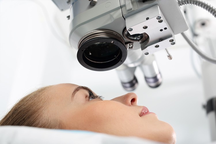
Diagnosis
The diagnosis of retinitis pigmentosa is based on a careful and detailed anamnesis, aimed at a thorough study of the present symptoms and the observation of other cases of retinitis pigmentosa in the family.
The presence of visual field narrowing and problems with night-blindness, lesions and pigment deposits in the eye fundus test is assessed.
All essential steps to rule out other eye diseases that share the same symptoms.
The diagnostic pathway makes use of specific tests:
- The electroretinogram (ERG) uses the same operation as an ECG, but with the substantial difference of focusing on studying the electrical stimuli produced by the retina. The ERG is useful for recording how the retina’s normal activity of collecting light stimuli is carried out and how it sends them to the occipital cortex for interpretation. The wave pattern is observed to see if the cells and various retinal components are responding correctly. Typically, in subjects with retinitis pigmentosa, the signal is reduced or even absent. The ERG is a useful test for all the patient’s family members to rule out or detect the disease at an early stage.
- The eye fundus test involves direct observation of the retina with a slit lamp, in order to show any granular abnormalities, pigmentary deposits at the back of the eye, narrowing of the retinal capillaries and extension of the spot obscuring vision.
- Optical Coherence Tomography (OCT) allows the different retinal layers to be assessed. This should show the lack of cones and rods, typical of retinitis pigmentosa. It is useful to highlight the stage of progression of the disease and to assess possible complications to the macula and vitreous.
- Given the strong hereditary component of the disease, a specialist examination of all family members is recommended, which may be followed by a genetic investigation to assess any alterations in the genes responsible for the onset of the disease.
Retinitis pigmentosa is a progressively progressive disease
It cannot be cured definitively, but it is possible to slow down its course.
In patients who have experienced complete loss of portions of the visual field, total recovery is impossible.
The disease is being studied and different therapeutic approaches are being tested.
Genetic analyses are essential in order to identify which genes are responsible for the onset of the disease, so that a pathophysiological basis can be established on which to develop a gene therapy, aimed at replacing the altered genes responsible for the disease with healthy ones.

New approaches, currently being tested, include the use of stem cells and the implantation of retinal prostheses, the latter being ideal for patients with advanced stages of the disease.
The ophthalmologist may find retinal or vitreous injections useful (certain active ingredients are inoculated to increase the proliferation of the remaining healthy cells).
Finally, hyperbaric oxygen therapy is the most useful therapy for slowing down the loss of photoreceptors, thus slowing down the progression of the disease.
Being a genetically determined disease, in most cases, there is no effective primary prevention.
To reduce the rate of complications and slow down the progression of the disease, exogenous intake of Vitamin A, Omega 3, lutein and zeaxanthin may be helpful.
Read Also
Emergency Live Even More…Live: Download The New Free App Of Your Newspaper For IOS And Android
What Is Presbyopia And When Does It Occur?
False Myths About Presbyopia: Let’s Clear The Air
Droopy Eyelids: How To Cure Eyelid Ptosis?
What Is Ocular Pterygium And When Surgery Is Necessary
Tear Film Dysfunction Syndrome, The Other Name For Dry Eye Syndrome
Vitreous Detachment: What It Is, What Consequences It Has
Macular Degeneration: What It Is, Symptoms, Causes, Treatment
Conjunctivitis: What It Is, Symptoms And Treatment
How To Cure Allergic Conjunctivitis And Reduce Clinical Signs: The Tacrolimus Study
Diabetic Retinopathy: The Importance Of Screening
Diabetic Retinopathy: Prevention And Controls To Avoid Complications
Diagnosis Of Diabetes: Why It Often Arrives Late
Diabetic Microangiopathy: What It Is And How To Treat It
Diabetes: Doing Sport Helps Blood Glucose Control
Type 2 Diabetes: New Drugs For A Personalised Treatment Approach
Diabetes And Christmas: 9 Tips For Living And Surviving The Festive Season
Diabetes Mellitus, An Overview
Diabetes, Everything You Need To Know
Type 1 Diabetes Mellitus: Symptoms, Diet And Treatment
Type 2 Diabetes Mellitus: Symptoms And Diet
Semaglutide For Obesity? Let’s See What The Anti-Diabetic Drug Is And How It Works
Italy: Semaglutide, Used For Type 2 Diabetes, Is In Short Supply
Gestational Diabetes, What It Is And How To Deal With It
Diabesity: What It Is, What Risks And How To Prevent It
Wounds And Diabetes: Manage And Accelerate Healing
The Diabetic Diet: 3 False Myths To Dispel
Top 5 Warning Signs Of Diabetes
Signs Of Diabetes: What To Look Out For