
Heart MRI and CT scan, what they are for
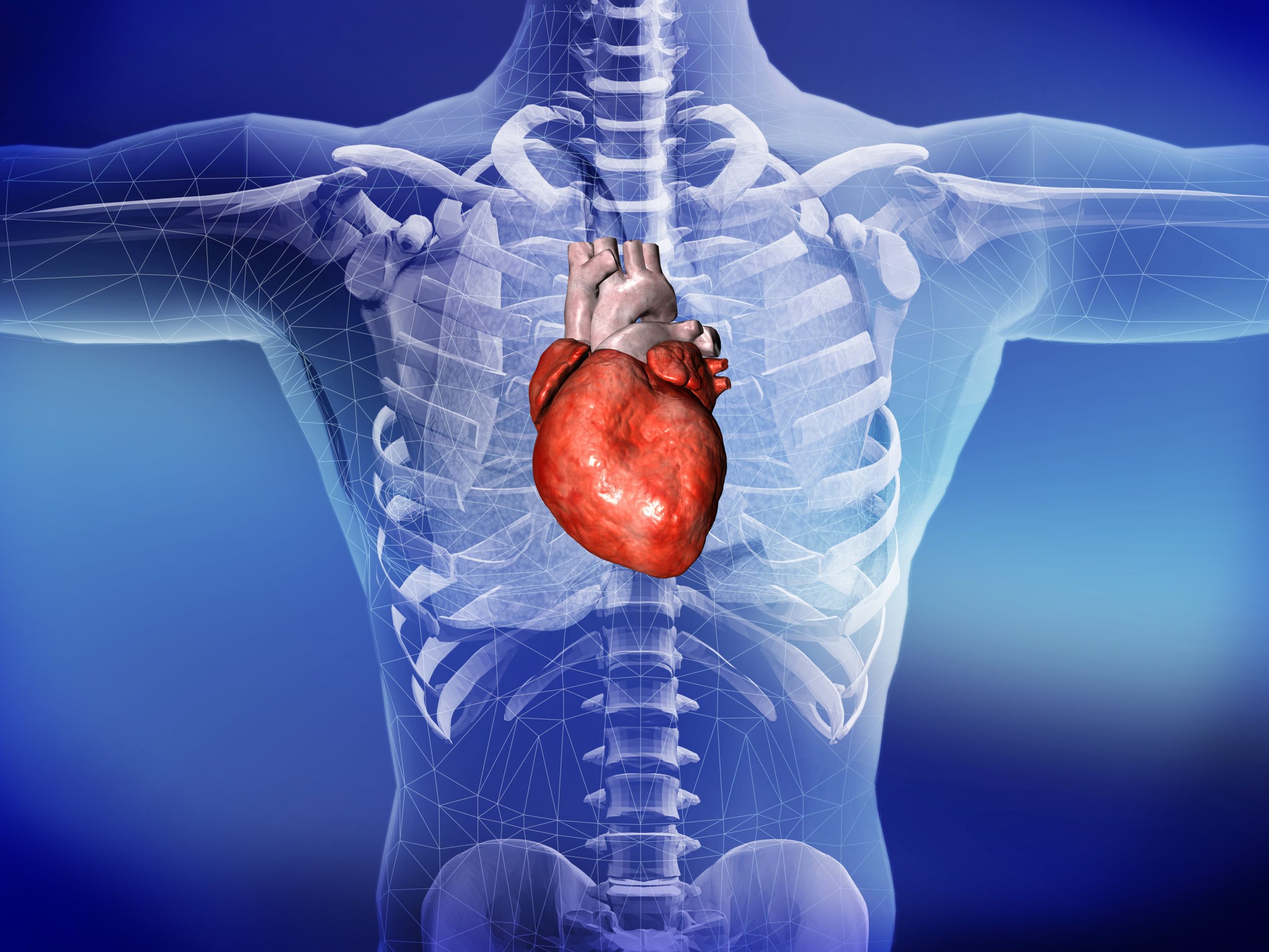
The heart is a complex organ, a structure in constant motion, with electrical activity and endowed with structures that are minute in size but fundamental in human physiology, such as the coronary arteries
Observing the heart requires the help of advanced technologies, which have been available for relatively few years and for which an interdisciplinary approach is required.
The use of these technologies requires not only cardiologists, but also radiologists to analyse the images and physicists to assess the technical and contractility aspects.
And we also need to train the specialists of tomorrow, so that they can keep up with technological developments.
Two fundamental examinations for cardiovascular health are the CT scan of the heart and the magnetic resonance imaging of the heart (MRI heart), let’s see what they are for and how they work.
Coronary arteries: what is a CT heart scan used for?
The CT scan of the heart is used to analyse the state of the coronary arteries.
It is a very fast examination: we are talking about a single beat acquisition, so with the latest generation machines it takes only three to four seconds to get a picture of the heart and arteries.
Thanks to the CT scan, one can assess the presence of plaques causing narrowing (stenosis) in the arteries, but also predict the risk of heart attack.
It is a fundamental prevention tool recognised by all international guidelines.
The CT scan is not an invasive examination, it emits some radiation but it is kept to a minimum, and it is generally painless.
Heart health: what MRI of the heart is for
Magnetic resonance imaging of the heart is an equally important tool that assesses the state of the heart muscle.
In fact, the heart can be affected by a wide variety of pathologies: from heart attacks to arrhythmias and myocarditis.
The examination lasts about 40 minutes, is non-invasive, does not emit X-rays, and allows the specialist to make a diagnosis beyond clinical evaluations.
MRI of the heart is a second-level examination that is essential for evaluating most pathologies affecting the heart and heart valves, such as:
- ischaemic heart disease
- dilated cardiomyopathies
- myocarditis
- hypertrophic cardiomyopathies
- congenital heart disease
- valvulopathies
- pericardial diseases.

It can also be used for the examination of vascular pathologies.
MRI of the heart, thanks to contrast medium, is the only method that makes it possible to visualise the presence of recent or previous structural damage to the heart: an infarction, myocarditis, inflammation of the pericardium, etc.
The examination therefore provides essential information for diagnosis and prognostic stratification in heart patients.
Heart and sport: symptoms not to be underestimated
Sporting activity is crucial in reducing the risk factors of several diseases and improving the prognosis of others.
If, however, those who do sport competitively follow rigorous fitness checks with very advanced examinations, those who do amateur sport to keep fit must pay attention to their body’s signals and if, while doing the same performance as always, they notice any different symptoms, they should consult a cardiologist.
Factors to watch out for are breathlessness, heart rate, which is normal and increases during physical activity but must not become excessively altered, or chest pains: these are all alarm bells to look out for.
In general, however, it is a good idea for those who exercise to undergo regular check-ups.
Read Also:
Emergency Live Even More…Live: Download The New Free App Of Your Newspaper For IOS And Android
What Is An ECG And When To Do An Electrocardiogram
Inflammations Of The Heart: Myocarditis, Infective Endocarditis And Pericarditis
MRI, Magnetic Resonance Imaging Of The Heart: What Is It And Why Is It Important?
CT, MRI And PET Scans: What Are They For?
Stroke, A Small CT Scanner On Ambulances And Helicopters To Aid Rapid Diagnosis