
High ferritin, low ferritin, normal values, significance, treatment: an overview
Ferritin is a globular protein found mainly in liver, spleen, bone marrow and skeletal tissue
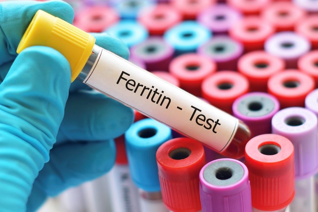
We also find small amounts of ferritin in plasma, which can be assessed by the so-called ferritinemia assay, which is obtained by means of a simple venous blood test.
Unlike iron bound to a second organic iron-protein compound called haemosiderin, the iron associated with ferritin is rapidly mobilised, meaning that in the event of a need for the mineral, the body can easily draw on it.
Ferritin is a 13 nm-diameter globular protein with a 6 nm core in which iron is contained
Within the pore structure of ferritin, iron ions are trapped and form the mineral ferridrite [FeO(OH)]8[FeO(H2PO4)] along with phosphate and hydroxide ions.
In ferritin, the iron is enclosed within a protein shell, the apopherritin, which can take up Fe2+ (ferrous ion) and oxidise it so that it is deposited as Fe3+(ferric ion).
Some forms of ferritin in vertebrates are hetero-oligomers of two highly related gene products with slightly different physiological properties.
The ratio of the two homologous proteins as a whole depends on the relative level of expression of the two genes.
The subunits that make up the structure have a molecular weight of 19 KDa (light chain L) and 21 KDa (heavy chain H).
The ratio between the amount of H and L chains varies depending on the tissue from which the ferritin originates.
The preponderance of L-light chains is typical of macromolecules with a large storage function, while those with a preponderance of H-chains have a greater capacity for buffering free radicals with consequent limitation of intracellular damage.
Ferritin can be glycosylated before being released into the circulation (GF)
Generally, between 50 and 80 per cent of the glycosylated portion is present in the circulation.
Function
Ferritin is the main storage protein for iron within cells, so its concentration in the blood reflects the extent of the mineral’s stores in the body.
Ferritin is mainly contained in cells, where it stores iron and releases it when needed, making it quickly available for use by the body.
A small proportion of ferritin is also transiently present in the bloodstream: this proportion is generally proportional to the concentration of the protein in the tissue.
Under normal conditions, there is a precise balance between the amount of ferritin present in the various tissues (stores) and plasma ferritin (circulating).
The concentration of the protein in the blood, although transient, is therefore an excellent indicator of the amount of iron available to the body.
Why is it useful to know ferritinemia?
Ferritinemia (i.e. the concentration of plasma ferritin) is very useful for assessing the amount of iron available to the body.
An abnormal level of ferritin in the blood can be an indicator of an underlying disease or a particular condition, as in the case of deficiencies responsible for anaemia.
In clinical practice, ferritinemia is generally assessed together with
- transferrinemia: the concentration of transferrin, the main iron transport protein in the blood;
- sideremia: the proportion of circulating transferrin that is saturated with iron;
- total iron binding capacity (TIBC): an indirect measure of transferrin’s ability to bind iron.
Normal ferritin values
Normal ferritinemia values vary according to the age and sex of the patient.
As a rule, ferritin values are slightly higher at birth; newborns may have values between 25 and 200 µg/ml, which may rise as early as the first month to 600 µg/ml, and then fall again during adolescence.
In adults, normal ferritinemia values are:
- women: 20-120 µg/mL;
- men: 20-200 µg/mL.
Causes of increased ferritinemia (hyperferritinemia)
An increase in serum ferritin indicates iron overload and can occur in various conditions and diseases:

- chronic alcoholism
- hepatocellular necrosis
- excessive dietary intake (e.g. excessive red meat);
- excessive use of drugs or iron supplements;
- haemochromatosis;
- hemosiderosis;
- chronic infections;
- leukaemias;
- cancer of the liver, lung, pancreas, breast and kidney;
- transfusions;
- Hodgkin’s lymphoma;
- acute or chronic hepatitis.
Causes of decreased ferritinemia (hypoferritinemia)
A decrease in serum ferritin indicates that iron reserves are low and can occur in various conditions and diseases:
- sideropenic anaemia (from iron deficiency);
- acute and chronic haemolytic anaemia;
- malnutrition;
- diet with iron-poor foods;
- reduced absorption
- vegetarian diet
- celiac disease
- chronic diarrhoea
- gastro-intestinal changes;
- haemorrhages of various kinds: from trauma, heavy menstrual flow, chronically bleeding haemorrhoids, occult haemorrhages
- pregnancy;
- rheumatoid arthritis.
Symptoms associated with low ferritinemia
Symptoms associated with iron deficiency anaemia are:
- feeling of severe tiredness and weakness;
- dyspnoea (difficulty breathing);
- difficulty in performing even mild exercises;
- brittleness of the nails;
- tachycardia (increased heart rate);
- tachypnoea (increased respiratory rate);
- dizziness;
- difficulty concentrating;
- increased thirst;
- blurred vision;
- splenomegaly (increased spleen volume);
- pain in the spleen (left flank);
- claudicatio intermittens: difficulty walking;
- confusional state;
- feeling of fainting;
- low body heat, especially in the extremities (hands and feet);
- noticeably pale appearance of the skin.
Therapy for altered blood ferritin values
In the case of altered blood ferritin levels, treatment must be based on the underlying cause.
Iron-rich foods
Here is a list of various iron-rich foods (iron value expressed per 100 grams of product):
- Goose liver 30.53 mg
- Bitter dark chocolate 17.4 mg
- Clam 13.98 mg
- Bitter cocoa 13,86 mg
- Cooked oyster 11,99 mg
- Caviar 11,88 mg
- Canned chicken pate 9.19 mg
- Muesli with fruit and dried fruit 8.75 mg
- Muesli 8.20 mg
- Lentils 7.54 mg
- Oyster 6,66 mg
- Soya flour 6,37 mg
- Wheat germ 6,26 mg
- Chicken (leg) 6,25 mg
- Chickpeas 6,24 mg
- Boiled potatoes 6,07 mg
- Cuttlefish 6,02 mg
- Dried pine nuts 5,53 mg
- Cannellini beans 5,49 mg
- Fresh borlotti beans 5,00 mg
- Oat flakes 4,72 mg
- Hazelnuts 4,70 mg
- Anchovies in oil 4.63 mg
- Peanuts 4,58 mg
- Durum wheat 4,56 mg
- Dried almonds 4,51 mg
- Hazelnut and cocoa cream 4,38 mg
Read Also
Emergency Live Even More…Live: Download The New Free App Of Your Newspaper For IOS And Android
Iron Deficiency Anaemia: What Foods Are Recommended
Iron, Ferritin And Transferrin: Normal Values
Increased ESR: What Does An Increase In The Patient’s Erythrocyte Sedimentation Rate Tell Us?
Anaemia, Vitamin Deficiency Among Causes
Mediterranean Anaemia: Diagnosis With A Blood Test
Colour Changes In The Urine: When To Consult A Doctor
Why Are There Leukocytes In My Urine?
How Iron Deficiency Anemia (IDA) Is Treated
Mediterranean Anaemia: Diagnosis With A Blood Test
Iron Deficiency Anaemia: What Foods Are Recommended
What Is Albumin And Why Is The Test Performed To Quantify Blood Albumin Values?
What Is Cholesterol And Why Is It Tested To Quantify The Level Of (Total) Cholesterol In The Blood?
Gestational Diabetes, What It Is And How To Deal With It