
Interventional radiology: what it is, why it is essential in patient care
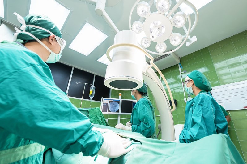
Interventional radiology: different instruments are used to obtain images of certain areas of the body, useful for making diagnoses and performing guided therapeutic interventions
Interventional radiology is a subspecialty of radiology
Doctors use different types of equipment to obtain images of certain areas of the body and use these images to perform image-guided interventions.
These imaging techniques include:
- Ultrasound;
- X-ray fluoroscopy;
- Computed tomography (CT);
- Magnetic resonance imaging (MRI).
Vascular interventional radiology provides extremely useful images for diagnosis and therapeutic intervention for a wide range of medical conditions
This procedure, which is relatively simple in the adult population, becomes progressively more complex as the size of the patient decreases.
Small catheters (small plastic tubes), usually 1-2 millimetres in diameter, are threaded through blood vessels to reach the site of the disease.
The catheter may be used to inject drugs, nutrients or hemodialysis into the vein.
In other cases, the catheter may be fitted at the tip of various instruments:
- To take blood samples for examination in the laboratory;
- To take images that are transmitted to a screen;
- To measure blood pressure;
- To take a tissue sample (biopsy);
- To inject a substance or contrast agent that is visible on X-rays (angiography);
- To widen a narrowed (stenosis) or blocked blood vessel (angioplasty);
- To widen the opening of a heart valve (valvuloplasty);
- To introduce substances or materials capable of occluding a blood vessel (embolisation).
The procedures for inserting a catheter vary greatly depending on the examination or therapy.
Some require general anaesthesia, others can be done under local anaesthesia.
In children, much of the interventional radiology work is directed towards obtaining medium to long-term vascular access, which is typically achieved with a central venous catheter or CVC
The main indications for the placement of a central venous catheter include the need for total parenteral nutrition, haemodialysis, intravenous administration over long periods of time of certain drugs including chemotherapy and antibiotics.
Another key indication in children is cardiac catheterisation for the diagnosis of congenital heart disease and, in many cases, for its treatment.
Arterial procedures have many of the same indications as in the adult population, such as embolisation of an arterial haemorrhage.
Treatment of some vascular malformations can be performed in childhood, thus reducing the symptoms and psychological problems associated with them.
The most common indication for renal angiography is renal hypertension.
Up to 25% of children and young people with hypertension have a renal cause. Stenosis (narrowing) of the renal artery, particularly due to fibromuscular dysplasia and neurofibromatosis type 1, can be successfully treated with angioplasty.
Angioplasty involves inserting a balloon catheter into a large blood vessel, e.g. the femoral artery, and pushing it until it reaches the narrowing.
The balloon is then inflated to widen the narrowing of the renal artery.

Diagnostic cerebral angiography has largely been replaced by imaging modalities such as CT angiography and MR angiography.
The diagnosis of some diseases, however, requires evaluation of small vessels and cerebral angiography often provides the resolution that is needed to diagnose, especially in younger children:
- Vasculitis;
- Spinal arteriovenous shunts;
- Cerebral aneurysms;
- Arteriovenous malformations.
In addition, a variety of interventions are now common including:
- The treatment of vascular lesions, such as aneurysms and arteriovenous malformations;
- The preoperative embolisation of tumours;
- The treatment of acute ischaemic stroke.
Embolisation involves the introduction, through catheters inserted into blood vessels, of materials capable of occluding a vessel such as surgical glue or metal coils.
Embolisation of the Galen vein malformation is a very successful procedure that has been shown to lead to neurological normalisation in 74% of treated patients.
In trauma, splenic embolisation is recognised by surgeons as an option for treating traumatic haemorrhage of the spleen.
Embolization of the splenic artery – the main blood vessel carrying blood to the spleen – allows the artery to be occluded and the bleeding to stop.
However, the spleen remains in place, continues to function at least partially, and avoids the risk of serious infections that patients who have had their spleen surgically removed (splenectomised) run.
Infections, cystic fibrosis, bronchiectasis, tuberculosis, foreign body aspiration and congenital heart disease can cause pulmonary haemorrhages to such an extent that embolisation of the bronchial arteries is necessary.
Blood vessel occlusions are possible complications of liver transplantation.
Angioplasty is the treatment of choice for hepatic arterial stenosis and both hepatic and portal venous stenosis.
Percutaneous sclerotherapy involves injecting a substance into a vein that destroys the blood vessel.
It is considered a first-line treatment for some venous malformations and is performed under fluoroscopic or ultrasound guidance.
Read Also:
Emergency Live Even More…Live: Download The New Free App Of Your Newspaper For IOS And Android
Chest Pain In Children: How To Assess It, What Causes It
Bronchoscopy: Ambu Set New Standards For Single-Use Endoscope
Bronchiectasis: How To Recognise And Treat It