

Lymphangiomas and lymphatic malformations: what they are, how to treat them
Lymphatic malformations, also known as lymphangiomas, are a group of diseases characterized by poor or absent development (hypo or aplasia) of lymphatic vessels and alterations in the circulation of lymphatic fluids
Depending on their characteristics, lesions are divided into microcystic and macrocystic or mixed.
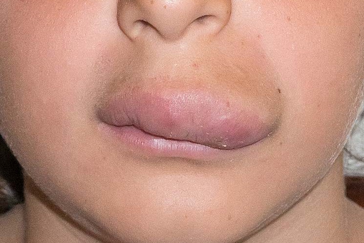
Lymphangiomas occur mainly during childhood and almost 50% are located in the head and neck
Growth is slow and distribution is local or diffuse.
In fortunately rare forms they may involve skin and subcutis, infiltrating muscle tissue and internal organs.
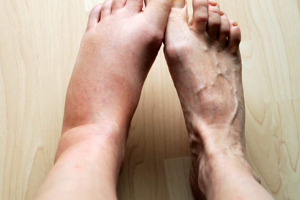
Macrocystic lymphatic malformations are relatively common, not only in the limbs, but also around the eye sockets, neck, tongue and floor of the mouth with protruding soft masses.
Microcystic malformations often manifest as multiple skin vesicles or warty nodules that secrete an exudate containing red blood cell breakdown products (serum fluid).
Thickening of the stratum corneum of the epidermis (cutaneous hyperkeratosis) is common.
The most common complications – occurring in untreated lesions – are infection and bleeding; in the case of bleeding, (intracystic microhemorrhage) the volume of the lesion suddenly increases and bluish infiltrations of blood appear under the skin (ecchymoses).
Symptoms of lymphangiomas
Lymphatic malformations that extensively affect a limb may be accompanied by swelling (lymphoedema) and skin imperfections.
These conditions may be accompanied by alterations in serum protein electrophoresis in laboratory tests.
Usually, lymphatic malformations and venous blood vessel malformations coexist, so we speak of capillary-lymphatic malformations or lymphatic-venous malformations, depending on the prevalent malformed tissue.
Lymphangiomas, diagnosis
The causes of lymphatic malformations remain unknown.

Diagnosis is based on medical examination, ultrasound and MRI, which is the most important diagnostic test as it allows a fairly precise estimate of the location, extent and anatomical structures involved.
With direct puncture, a yellowish liquid can be aspirated and, at the same time, sclerotherapy can be performed in preparation for surgical removal.
How lymphatic malformations are treated
Most lymphatic malformations of the hand are microcystic.
This means that there is little scope for sclerosing therapy.
The treatment today consists mainly of sclerosant therapy or sclerotherapy and surgical resection, which is reserved for microcystic forms.
The goal of completely removing a lymphatic malformation of the hand is unfortunately optimistic, because recurrences can always occur, even years later.
Sclerotherapy is a procedure that consists of injecting a compound into the malformed vessel(s), which can cause them to close and collapse.
The solution containing the compound is called “sclerosing solution”.
Surgical therapy is always approached in stages, with frequent recurrences due to the extensive spread of lymphatic tissue.
Read Also:
Emergency Live Even More…Live: Download The New Free App Of Your Newspaper For IOS And Android
Lymphoma: 10 Alarm Bells Not To Be Underestimated
Non-Hodgkin’s Lymphoma: Symptoms, Diagnosis And Treatment Of A Heterogeneous Group Of Tumours
CAR-T: An Innovative Therapy For Lymphomas
Acute Lymphoblastic Leukaemia: Long-Term Outcomes Described For Childhood ALL Survivors