Retinal detachment: what is it?
Retinal detachment is one of the most frequent diseases in ophthalmology, and often has a traumatic origin: an injury in the affected area of the face causes this serious medical problem
The condition of retinal detachment occurs when the inner membrane of the eye – the retina, a thin layer of tissue lining the back of the eye – detaches from the supporting tissues.
Depending on the cause of the retinal detachment, this disease is classified into three different types: rhegmatogenous, tractional and exudative.
Rhegmatogenous retinal detachment
Retinal detachment, if rhegmatogenous, is determined by the initial and progressive degeneration of the retina.
As it degenerates, it loses its biological adherence to the pigment epithelium and – in the actual retinal ruptures or tears that occur – the vitreous body, a gelatinous and transparent fluid that fills the interstice between the posterior part of the crystalline lens and the retinal wall, causes it to detach.
Tractional retinal detachment
In this case there is the formation of a pathological fibro-vascular tissue that tends to wrinkle and exert traction on the retinal surface causing it to detach.
Certain diseases predispose to the formation of this pathological tissue, in particular proliferative diabetic retinopathy.
Exudative retinal detachment
Caused by retinal disease without tears, inflammatory disorders, traumatic events or vascular abnormalities, exudative retinal detachment occurs when an abnormal amount of fluid accumulates between the retina and its supporting tissues.
Various causes behind retinal detachment
Some of the most common include:
Ageing
Advancing age is one of the most common causes of retinal detachment, vitreous detachment and retinal rupture.
Generally, ageing is also associated with severe myopia (greater than 5 or 6 dioptry), which can contribute to retinal detachment – which is already very thinned – also of a spontaneous type due to the structural characteristics typical of the strongly myopic eye.
Traumatic events
Retinal detachment, in some cases, can be caused by traumatic events occurring to the face or the eyeball itself: a penetrating injury, for example. Generally, traumatic events triggering retinal detachment are associated with the practice of contact or combat sports or accidents occurring at high speed.
Surgical complications
Some surgical procedures performed on the eye can have retinal detachment as one of their complications.
In fact, a retina with areas of weakness can become detached even after cataract removal surgery.
Diabetes
Retinal detachment may be counted among the complications of diabetes.
In such a case, the pathology from which the eye suffers is called diabetic retinopathy and may result in tractional retinal detachment, or due to intense neovascularisation and microvascular changes to the retinal tissue.
Inflammatory diseases
Some inflammatory diseases affecting the eye – such as uveitis or melanoma of the choroid – can cause localised inflammation and intraocular swelling, resulting in detachment of the retina, which remains intact, from its supporting tissues and infiltration of the remaining empty space by exudative fluids.
However, it remains important to specify that retinal detachment is a rather rare condition.
It is most likely to occur in adult and elderly patients between 50 and 75 years of age; if retinal detachment occurs while still young, the causes will almost exclusively be found in traumatic events.
Retinal detachment – the symptoms
Retinal detachment is a serious condition whose symptoms begin to be felt immediately and strongly by the patient.
It is not associated with any painful symptoms.
Once the detachment has occurred, the first symptom felt will be myodesopias: a sudden appearance of small black dots, dark spots or floating streaks within the visual field.
Some patients describe a kind of ‘spider’s web effect’, while others report the presence of a single large black dot.
Subsequently, the retinal detachment patient may complain of the appearance of photopsia: real flashes of light perceived at the periphery of the visual field of the affected eye.
Accompanying the photopsia, there may also be phenomena of blurred or distorted vision.
In addition, it is accompanied by an amputation of the visual field, i.e. the patient perceives a kind of ‘black curtain’ that absolutely prevents vision in a portion of the visual field
As mentioned above, retinal detachment is a serious phenomenon that must be treated promptly.
If the symptoms are ignored or underestimated, vision will slowly deteriorate and any treatment options will be less effective.
Diagnosing retinal detachment
The earlier the patient who has detected the previous symptoms decides to visit an eye doctor for a specialist consultation, the greater the chances of regaining good function of the retinal detachment eye.
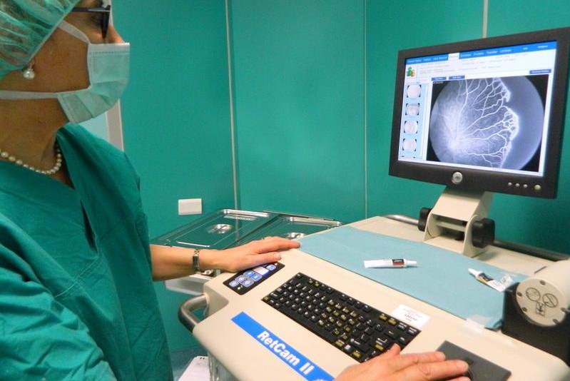
During the specialist consultation, the ophthalmologist – after taking a thorough anamnesis – will immediately proceed with the assessment of the integrity of the posterior portion of the eye.
This will be done by means of ophthalmoscopy, a specialised test that uses an instrument that – by projecting a beam of light through the pupil onto the retina – is able to provide information about the internal structures of the patient’s eye, especially if these structures are altered, torn or damaged.
The ophthalmologist may also use the slit lamp to test the anterior and posterior structures of the eye.
Retinal detachment: the most appropriate therapy
Once the ophthalmologist has completed all the tests described above, he can formulate the precise diagnosis of retinal detachment.
Based on the findings, the patient will be told the most appropriate therapy to follow. In the case of retinal detachment, the treatment is surgical.
There are different surgical approaches depending on the extent of the retinal detachment.
Laser surgery is indicated for correcting small ruptures and preventing evolution into a massive retinal detachment.
In the case of a retinal detachment that has occurred, there are various techniques such as pneumoretinopexy, episcleral surgery using a scleral buckle and vitrectomy.
The choice of the most suitable surgery lies exclusively with the ophthalmologist who will decide on the most suitable treatment for the patient, and in some cases multiple surgeries will be necessary.
Often eye function does not return as it originally was, as recovery of normal visual capacity depends on both the extent and type of the retinal detachment.
However, treatment carried out early and with the right clinical indication guarantees a good recovery of visual function.
Read Also
Emergency Live Even More…Live: Download The New Free App Of Your Newspaper For IOS And Android
Retinal Detachment: Symptoms And Causes
Heart Attack, Prediction And Prevention Thanks To Retinal Vessels And Artificial Intelligence
Symptoms, Causes And Treatment Of Dacryocystitis
Entropion: The Symptoms And How To Treat It
Dacryocystitis: Definition, Symptoms, Causes, Diagnosis And Treatment
Early Diagnosis Of Maculopathies: The Role Of Optical Coherence Tomography OCT
What Is Maculopathy, Or Macular Degeneration
Inflammations Of The Eye: Uveitis
Corneal Keratoconus, Corneal Cross-Linking UVA Treatment
Myopia: What It Is And How To Treat It
Presbyopia: What Are The Symptoms And How To Correct It
Nearsightedness: What It Myopia And How To Correct It
Blepharoptosis: Getting To Know Eyelid Drooping
Lazy Eye: How To Recognise And Treat Amblyopia?
What Is Presbyopia And When Does It Occur?
Presbyopia: An Age-Related Visual Disorder
Blepharoptosis: Getting To Know Eyelid Drooping
Rare Diseases: Von Hippel-Lindau Syndrome
Rare Diseases: Septo-Optic Dysplasia
Diseases Of The Cornea: Keratitis
Cataract: Symptoms, Causes And Intervention
Eye For Health: Cataract Surgery With Intraocular Lenses To Correct Visual Defects


