
Soft tissue tumours: leiomyosarcoma
Leiomyosarcoma is a soft tissue tumour that mainly affects women and rarely children. Resistant to chemotherapy, surgery is the only treatment
Leiomyosarcoma is a malignant soft tissue tumour and belongs to the sarcoma family
It develops from smooth muscle cells.
It is an adult disease, with a peak incidence in the 5th and 6th decade of life and a predilection for the female gender, which, albeit rarely, can also affect children.
In paediatric age, boys and girls are equally affected.
Leiomyosarcomas are subdivided into five groups based on the mode of presentation:
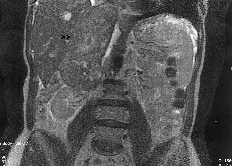
- “Retroperitoneal soft tissue leiomyosarcoma”;
- “Leiomyosarcoma of cutaneous origin”;
- “Leiomyosarcoma of vascular origin”:
- “Leiomyosarcoma in immunocompromised patients”;
- “Leiomyosarcoma of bone”.
The most frequent site of development (50% of cases) is the retroperitoneum.
The cause of occurrence of this tumour is unknown and no relationship with environmental causes has been established.
Patients with hereditary retinoblastoma, carriers of the constitutional mutation of the Rb1 gene, have an increased risk of developing leiomyosarcoma in adulthood compared to the general population.
The predilection for the female gender has also led to speculation that oestrogen plays a role in smooth muscle proliferation, however the correlation remains unclear.
As for Leiomyosarcoma in the immunocompromised patient, a correlation with the Epstein-Barr virus is usually found, and in these cases the tumour may arise in non-typical sites.
Leiomyosarcomas, like all other sarcomas, have non-specific presenting symptoms due to the presence of a lesion that compresses, dislocates and infiltrates surrounding structures.
The mode of presentation of leiomyosarcoma therefore varies depending on the site of onset
“Soft tissue leiomyosarcoma” usually manifests as a retroperitoneal lesion, in which case the presenting symptoms are vague abdominal discomfort, more or less associated with weight loss.
Leiomyosarcoma of cutaneous origin’ usually presents as a small intra-dermal swelling.
Leiomyosarcoma of vascular origin’ develops from large blood vessels and frequently affects the pulmonary artery.
In this case, the symptoms are mainly respiratory difficulty and chest pain due to obstruction of blood flow.
Leiomyosarcoma of the bones’ is a very rare entity and usually affects the long bones
Whenever the presence of a sarcoma is suspected, on the basis of the objective examination and preliminary radiological investigations, a fragment of the tumour must be taken and analysed by the pathologist (possibly an expert in this type of lesion), the specialist able to make the diagnosis.
There are also further investigations that assess the characteristics and extent of this tumour:
- Nuclear Magnetic Resonance Imaging (MRI) and/or Computed Tomography (CT), which allow the size of the tumour, its extension and relationship with adjacent structures to be assessed;
- Radiography or CT scan of the chest, plus or minus bone scintigraphy for evaluation of any distant secondary lesions.
- Surgery is the mainstay of treatment for leiomyosarcoma and, in cases where removal of the lesion is radical, may be the only treatment.
In cases where excision is incomplete, complementary local treatment with radiotherapy may be necessary, especially for lesions of the extremities.
Leiomyosarcoma is not a chemotherapy-sensitive tumour

The role of cytostatic treatment is unclear and consequently chemotherapy is reserved for selected cases: large lesions, inoperable in the first instance, with the aim of reducing tumour volume and associated symptoms; patients with lesions that recur at a distance.
The most commonly used chemotherapeutic agents are anthracyclines, ifosfamide, gemcitabine, taxotere, dacarbazine and trabectidine.
With regard to therapies targeting the tumour cell, of the various molecules tested, the only one that has shown moderate efficacy against leiomyosarcoma is pazopanib, a tyrosine kinase inhibitor, administered orally.
The prognosis of a patient with leiomyosarcoma depends on the histological features of the lesion, the local extension (size of the mass and relationship with surrounding structures) and the anatomical site of the lesion, as well as any distant recurrence.
If the entire tumour mass can be removed, the prognosis is very good with an 80% chance of cure.
Extremity tumours have a better prognosis than retroperitoneal tumours, as they are smaller lesions and the chance of radical surgical removal is higher.
Intradermal lesions have an excellent prognosis as they tend to remain localised and not metastasise.
The prognosis remains less favourable for patients with distant recurrent lesions.
Read Also
Emergency Live Even More…Live: Download The New Free App Of Your Newspaper For IOS And Android
Soft Tissue Sarcomas: Malignant Fibrous Histiocytoma
Brain Tumours: Symptoms, Classification, Diagnosis And Treatment
Pediatric Brain Tumors: Types, Causes, Diagnosis And Treatment
Brain Tumours: CAR-T Offers New Hope For Treating Inoperable Gliomas
Lymphoma: 10 Alarm Bells Not To Be Underestimated
Chemotherapy: What It Is And When It Is Performed
CAR-T: An Innovative Therapy For Lymphomas
What Is CAR-T And How Does CAR-T Work?
Radiotherapy: What It Is Used For And What The Effects Are
Craniosynostosis Surgery: Overview
Pediatric Malignancies: Medulloblastoma