
What is the full-spine radiography and what is it for?
Let’s talk about X-rays: the full-spine radiography of the spinal column is an examination that makes it possible to check the actual condition and state of health of the spinal column in just 10 minutes and detect any problems
What is the full-spine radiography for?
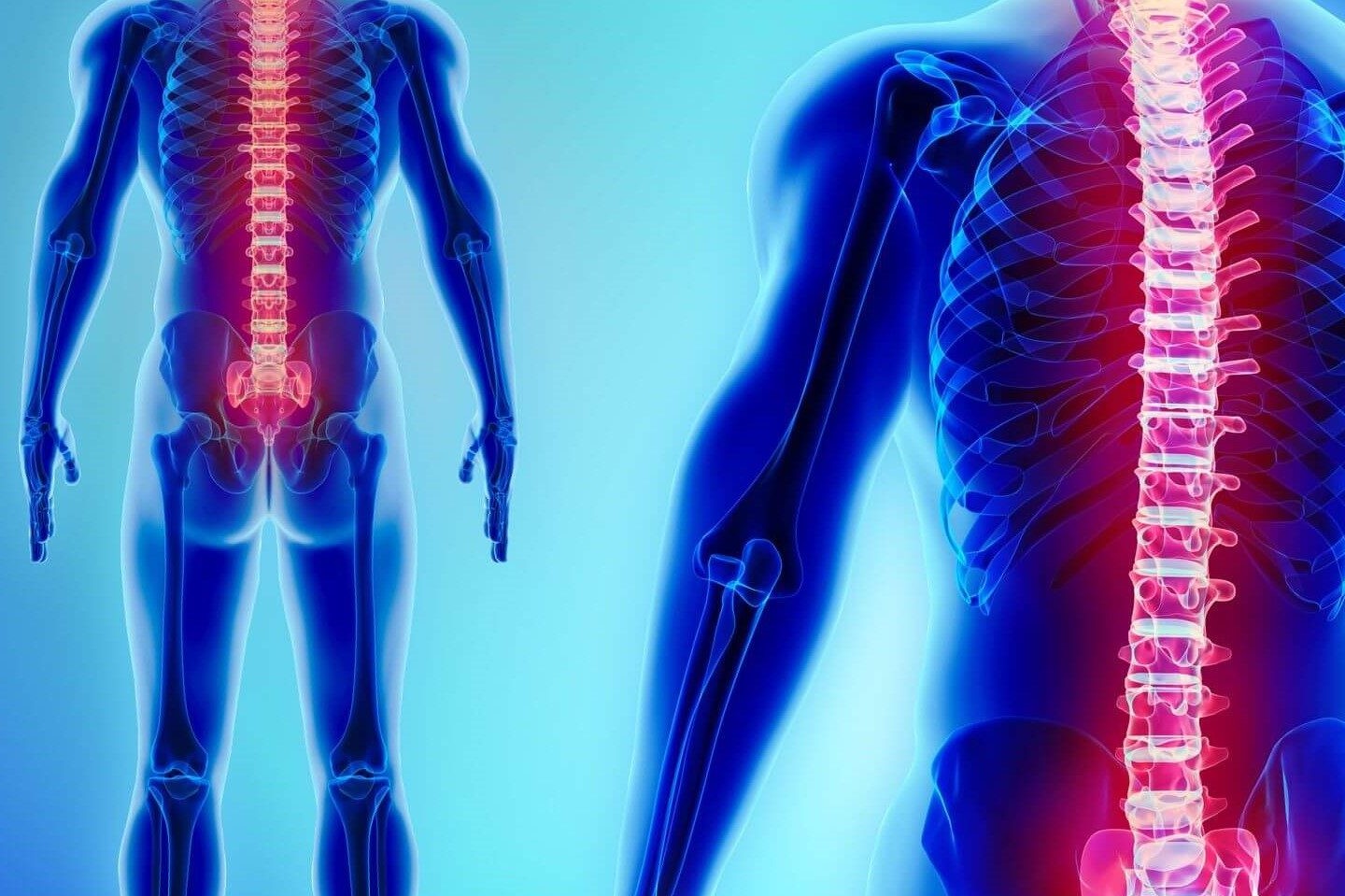
The full-spine radiographyof the spinal column, performed using X-rays, makes it possible to obtain in a single image a ‘snapshot’ of the entire spinal column, from the cervical to the pelvis, and to visualise and therefore assess
- the cervical tract
- the dorsal tract;
- the lumbosacral tract.
Does the X-ray hurt?
It is a safe and non-invasive examination. The technologies available today, in fact, while using X-rays, allow it to be performed in total safety: the doses of radiation currently used to perform the examination and the exposure time have been significantly reduced compared to past years.
The only contraindication is, as with other traditional X-ray investigations, pregnancy.
The full-spine radiography is a safe and reliable examination that allows the detection of
- congenital alterations (e.g. scoliosis, lordosis, kyphosis)
- acquired alterations;
- post-traumatic degenerative processes.
In addition to these deformations of the spinal column, the examination is able to detect abnormalities in the weight-bearing axes of the lower limbs and dysmetria of the pelvis due to scoliosis and/or the different length of the lower limbs.
Finally, it also allows the spinal column to be studied at the postural level.
How it is done
To perform this examination, which lasts approximately 10 minutes (except in special cases), the person is made to stand
- standing (in this case we talk about ‘loaded examination’) near the radiographic equipment;
- lying on a special bed, and is only asked to cooperate a little, in order to assume the necessary positions so that the examination can be carried out optimally.
Once the X-ray examination is complete, you can return to everyday life immediately.
Full-spine radiography: How to prepare for the examination
There are no special preparation rules.

However, it is advisable to wear comfortable clothes and avoid wearing metal objects that should be removed during the examination as they could interfere with obtaining the X-ray.
This X-ray may be requested by physiatrists, orthopaedists and paediatricians for the purpose of monitoring structural growth in particular
- in adolescents;
- in arthrosis sufferers with hip or knee replacements.
Read Also:
Emergency Live Even More…Live: Download The New Free App Of Your Newspaper For IOS And Android
Spinal Canal Decompression: What It Is And When It Is Performed
Spinal Immobilization: Treatment Or Injury?
10 Steps To Perform A Correct Spinal Immobilization Of A Trauma Patient
Spinal Column Injuries, The Value Of The Rock Pin / Rock Pin Max Spine Board
Spinal Immobilisation, One Of The Techniques The Rescuer Must Master
Spinal Immobilisation Of The Patient: When Should The Spine Board Be Put Aside?
Intraosseous Access, A Life-Saving Technique In Emergency Shock Management
Electromyography (EMG), What It Assesses And When It Is Done
Dislocation Of The Shoulder: How To Reduce It? An Overview Of The Main Techniques
What Is Hand Radiography (Hand X-Ray)?