
Congenital heart disease, a new technology for pulmonary valve prostheses: they are self-expanding via transcatheter
The Bambino Gesù is the European leader in the use of trans-catheter self-expanding pulmonary valve prostheses to treat congenital heart disease
Congenital heart disease, two boys have already been successfully treated
A self-expanding pulmonary valve prosthesis that can be adapted more effectively to the anatomy of children and young people with congenital heart disease: this is the innovative system offered by technology in the field of trans-catheter procedures, those that make it possible to avoid open-heart surgery.
The new prosthesis, which received EC authorisation in May this year, is already a reality in the Interventional Cardiology Unit of the Bambino Gesù Children’s Hospital, directed by Dr Gianfranco Butera, who has successfully used it for two boys aged 15 and 19.
QUALITY AED? VISIT THE ZOLL BOOTH AT EMERGENCY EXPO
PULMONARY VALVE DEFECTS
The pulmonary valve is one of the four cardiac valves: placed between the right ventricle and the pulmonary artery, it has the task of ensuring that the blood proceeds smoothly, without reflux, on its way to the lungs to replenish itself with oxygen.
Certain pathologies impair its functioning.
This is the case with tetralogy of Fallot, which is one of the most frequent severe congenital heart diseases.
Due to malfunctioning, the right ventricle is overworked and can decompensate with very serious consequences.
It is therefore often necessary to implant a pulmonary valve prosthesis to replace the damaged valve.
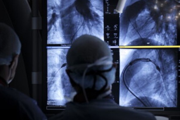
The procedure is performed in the haemodynamics room in a minimally invasive manner, without the need for open-heart surgery, by inserting a cannula into an artery, inside which a catheter (small flexible tube) of minimal size is run and positioned where the problem is to be solved.
CONGENITAL HEART DISEASE, ‘BALLOON EXPANDABLE’ TECHNOLOGY
Trans-catheter pulmonary valve prostheses have been in clinical practice since 2007.
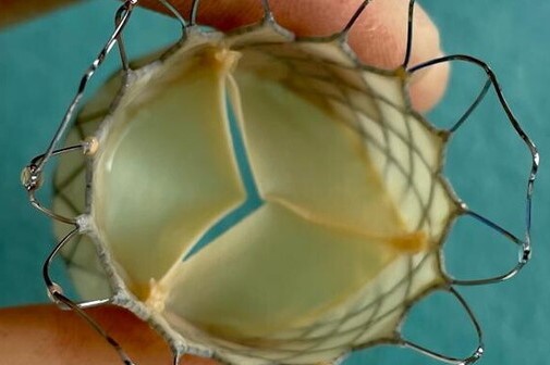
They are made of biological material (made from a pig or bovine valve analogue, treated and fixed on a metal ring covered with synthetic fibre) and are all based on ‘balloon expandable’ technology.
The valve is sewn inside a metal support (stent) that is implanted by means of a balloon introduced through the catheter.
The balloon is inflated and expands the stent, placing the prosthesis in the desired position.
The size of the balloon – approximately 16 to 29 millimetres – is, however, a limitation in using the system.
In most patients, in fact, the space in which to insert the valve prosthesis is larger, as a consequence of disease, previous operations or other events in the medical history.
This is the reason why with ‘balloon expandable’ technology it is only possible to treat about 20% of patients who need a pulmonary valve replacement.
THE NEW ‘SELF-EXPANDABLE VALVE’ TECHNOLOGY FOR THE TREATMENT OF CONGENITAL HEART DISEASE
The new technology is based on the ‘self-expandable valve’ system.
The stent is self-expandable and can reach larger diameters – up to about 36 millimetres – greatly increasing the number of patients eligible for treatment.
Approximately 30 children and young people are operated on each year at the Bambino Gesù: a number that can be more than doubled with the new technique by avoiding open-heart surgery.
Many of the patients have previously undergone surgery, so the possibility of access to a minimally invasive operation is an undeniable advantage.
The system led by the Holy See Hospital was developed by a Chinese company and received the CE mark in May 2022 (Venus Valve).
At present, there are no other similar systems that can be used in clinical practice.
The Bambino Gesù is the first European hospital to use this technology after clinical approval.
It has also been identified as a reference point for Italy and Europe and will be responsible for promoting its use in other centres, also taking care of operator training.
THE CHILDREN TREATED AT BAMBINO GESU
The first two patients treated at the Bambino Gesù, aged 15 and 19, had a previous history of multiple operations due to heart disease.

Further cardiac surgery would have represented a very significant risk.
In both cases, additional risk factors such as scoliosis, previous stroke, and impaired functional capacity were also present.
The collaboration between the interventional cardiology unit and the thoracic and advanced cardiovascular radiology unit headed by Dr Aurelio Secinaro is crucial for patient selection.
In addition to magnetic resonance imaging (MRI) and computed tomography (CT), which allow detailed information on the anatomy and function of the cardiovascular system to be obtained in a less invasive manner, the role of 3D technology is relevant.
Three-dimensional image reconstruction makes it possible to highlight the points of the heart wall and pulmonary artery on which the valve will rest and allows for an accurate assessment of patients eligible for treatment.
The computer simulation of the operation, moreover, thanks to an algorithm that processes the data of the specific boy, makes it possible to anticipate any problems that may occur and to better plan the procedure.
The two boys who received the pulmonary valve prosthesis were also spared a stay in intensive care.
In addition, whereas the average hospital stay in a conventional operation is around two weeks, in these cases the patients were discharged only three days after the operation.
Today, they are doing well and have successfully passed the first monitoring and control phases.
PROSPECTS FOR INTERVENTIONAL CARDIOLOGY
At the Bambino Gesù there are already other guys ready for the next intervention session.
“It is possible to foresee,” says Dr. Butera, “that in Italy at least a hundred patients a year will be able to benefit from this technology, with a considerable reduction in the impact on the National Health Service as well, but, above all, with a considerable reduction in physical and psychological pain and risks for our boys and great satisfaction for their families.
The prospect, Dr Butera continues, is certainly that of: “greatly expand the number of patients who can benefit from a minimally invasive approach.
Moreover, the application of imaging techniques, 3D visualisation, and computational simulation will allow us to understand even better the margins of application and to broaden the situations in which we can proceed’.
Read Also:
Emergency Live Even More…Live: Download The New Free App Of Your Newspaper For IOS And Android
Defibrillator: What It Is, How It Works, Price, Voltage, Manual And External
The Patient’s ECG: How To Read An Electrocardiogram In A Simple Way
Signs And Symptoms Of Sudden Cardiac Arrest: How To Tell If Someone Needs CPR
Inflammations Of The Heart: Myocarditis, Infective Endocarditis And Pericarditis
Quickly Finding – And Treating – The Cause Of A Stroke May Prevent More: New Guidelines
Atrial Fibrillation: Symptoms To Watch Out For
Wolff-Parkinson-White Syndrome: What It Is And How To Treat It
Do You Have Episodes Of Sudden Tachycardia? You May Suffer From Wolff-Parkinson-White Syndrome (WPW)
Transient Tachypnoea Of The Newborn: Overview Of Neonatal Wet Lung Syndrome
Tachycardia: Is There A Risk Of Arrhythmia? What Differences Exist Between The Two?
Bacterial Endocarditis: Prophylaxis In Children And Adults
Erectile Dysfunction And Cardiovascular Problems: What Is The Link?
Ischaemic Heart Disease: What It Is, How To Prevent It And How To Treat It
Ischaemic Heart Disease: Chronic, Definition, Symptoms, Consequences