
Bone callus and pseudoarthrosis, when the fracture does not heal: causes, diagnosis and treatment
When a bone suffers a fracture, under physiological conditions a biological repair process begins, which over time leads to the formation of ‘bone callus’.
The bone callus is a repair tissue that is created by the process of callogenesis that usually occurs three weeks after the traumatic event that led to the fracture
The bone callus welds the fragments of the fractured bone and gradually modulates in response to the mechanical forces exerted on it, becoming increasingly resistant.
In the following weeks or months, the bone callus reconstitutes the integrity and normal biomechanical characteristics of the injured skeletal segment, however – if the calcification process undergoes such a conditioning or interruption that it does not allow consolidation – the fracture may not heal properly.
In this case, a fibrous callus is formed, which leads to pain and functional limitation (pseudoarthrosis) and often necessitates surgery. In some cases, one can speak of ‘delayed consolidation’ when the bone begins to form a callus but takes longer than normal to complete healing.
Bone healing can be hindered by certain pre-existing risk factors such as metabolic diseases or cigarette smoking.
What factors can affect bone healing and bone callus formation?
Bone heals when the fracture is stable and has sufficient vascularisation for bone callus to form.
Proper nutrition plays an important role in bone healing.
- Stability, alignment, mutual contact of the parts, immobility: the most important rule is that when a bone breaks, the broken parts must be realigned and in contact and must not move until they heal as even small movements during the formation of the bone callus can disturb healing and create a pseudo-arthrosis. Some fractures can be stabilised simply with a plaster cast others require surgical treatment with a reduction and stabilisation through synthetic means such as plates, screws, nails or external fixators.
- Vascularisation: the blood supply is fundamental for the healing of a fracture as all the factors that are indispensable for the formation of the bone callus are transported through the blood
- Nutrition: having an adequate diet is important to facilitate bone healing through a healthy and balanced diet that includes calcium, protein, vitamin C and D is the basis for proper bone healing, dietary supplements that go beyond the daily requirements are not necessary (the rare exception is for severely malnourished patients with metabolic diseases or multi-organ damage, in which case the doctor can advise on the best dietary guidelines and possibly add dietary supplements).
Stages of reparative osteogenesis of fractures
In summary, the phases leading to healing of a fracture are:
- haematoma formation and organisation phase (= haemorrhagic transfusion);
- phase of tissue proliferation and differentiation in an osteogenetic sense (the haematoma cells at the fracture site differentiate into osteocytes);
- maturation phase (i.e. hardening, calcification of the callus) and then remodelling phase (i.e. remodelling of the callus that tends to make the actual fracture marks disappear).
Causes of pseudoarthrosis
Bone does not heal and goes into pseudoarthrosis when it lacks sufficient stability or blood flow is reduced, situations that can sometimes coexist.
A high-energy trauma such as a car accident, for example, can cause a serious injury that not only breaks the bone but also leads to compromised vascularisation due to injury to the surrounding soft tissue.
There are several risk factors that increase the likelihood of a fracture leading to pseudoarthrosis:
- tobacco or nicotine use inhibits fracture healing and increases the likelihood of pseudoarthrosis formation
- advanced age
- severe anaemia
- diabetes
- low vitamin D levels
- hypothyroidism
- poor or poor diet
- use of notorious drugs such as acetyl-salicylic acid, ibuprofen and cortisone (the doctor should be aware of the drugs taken by fracture patients in order to assess the possibility of discontinuing therapy during the fracture healing period)
- infections
- exposed fractures (when the bone has protruded from the skin)
- vascularisation impairment
Some bones, such as those of the foot, have intrinsic stability and an excellent blood supply, in which case they can heal even with non-surgical treatment and minimal stability.
In some bones, such as the head of the femur or the scaphoid of the wrist, the fracture causes an interruption of the vascularisation and consequently the risk of pseudoarthrosis is high.
Some bones, such as the tibia, have a moderate blood supply; high-energy trauma can impair the skin condition and promote pseudoarthrosis of the fracture in this district.
Symptoms of pseudoarthrosis
Pseudoarthrosis is usually painful and when it occurs, it arises after a period of well-being following treatment of the fracture, then pain begins months after the fracture and may be persistent for months or years, or it may begin when using the broken arm or leg or may be present even at rest.
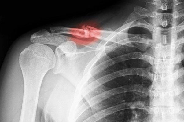
Diagnosis of pseudoarthrosis
To diagnose a pseudoarthrosis, the orthopaedist uses X-ray examinations, and depending on the district affected, simple X-rays or more specialised examinations such as CT or MRI may be required.
Through these investigations, the doctor determines the progress of healing or the presence of a pseudoarthrosis.

Pseudarthrosis is usually said to be present when the orthopaedic doctor finds from clinical and radiographic examinations
- persistent pain for more than 6 months at the fracture site
- a lack of bone callus formation within the appropriate biological time frame and during follow-up examinations in the following months
- resorption of the fracture stumps or a space between them
If pseudarthrosis is diagnosed, the doctor may ask for some blood tests to find out if there is a vitamin or calcium deficiency, a metabolic disorder such as diabetes and hypothyroidism, or if there is an infection.
Treatment of pseudarthrosis
Treatment can be surgical or non-surgical, and your orthopaedist will discuss with you the treatment options that best suit your case, outlining the risks and benefits of the choice to resolve the case of pseudoarthrosis.
1) Non-surgical treatment. The use of a bone stimulator such as magneto therapy or PEMF (pulsed electromagnetic fields) is applied to the skin in the area of pseudarthrosis, this small device delivers ultrasonic electromagnetic waves or pulses that stimulate bone healing. The device should be applied daily from 20 minutes to several hours depending on the instructions of your orthopaedist or physiatrist.
2) Surgical treatment. Surgery is necessary when traditional treatment methods fail. A new surgical treatment may be necessary if the first treatment did not heal the fracture. Surgical options include re-synthesis of the fracture, autologous or organ-donor bone grafting (allograft) or bone substitutes and internal and/or external synthesis.
- Autologous bone grafting: during this procedure, the orthopaedic surgeon takes bone from another district such as from the pelvis and places it at the site of pseudoarthrosis after removing pathological healing tissue from the fracture. The bone used has the function of functional and biological support, i.e. it serves to reinforce the stability of the synthesis and to supply cells and healing factors to the fracture site. The most commonly used site for bone harvesting is the pelvis, in which case the surgeon makes an incision at the edge of the iliac crest and from there will harvest enough bone tissue to treat the pseudarthrosis.
- Allograft (organ donor graft): an allograft avoids taking bone from the patient and thus reduces the duration of the operation and post-operative pain. It provides a scaffold and therefore functional support for fracture stability but gives no biological contribution as it is non-viable bone, which is why it is often used in combination with bone taken from the patient’s pelvis. Over time, allograft will either be resorbed or replaced by viable bone.
- Bone substitutes: as with allografts, bone substitutes have the advantage of shortening surgical times and reducing post-operative pain alone they do not provide functional or biological support they are treated with certain substances that activate and promote bone formation.
Very frequently the stability of the fracture in pseudoarthrosis is not provided by bone grafts but these must be associated with stabilisation through synthesis with internal fixators such as plates and screws or nails or external fixators:
- Internal fixation: If a pseudoarthrosis occurs after internal synthesis surgery, the surgical choice may be a new internal synthesis to increase stability. The surgeon may choose to replace an intramedullary nail with one of a larger diameter to increase the stability of the fracture and promote bleeding at the site of pseudoarthrosis or change a plate to increase stability by also using bone grafts to promote healing.
- External fixator is an external scaffold that is attached to the bone through rigid Fiches pins that are screwed into the bone itself away from the fracture and on these pins externally the scaffold is built to stabilise the fracture. External fixation can also be used in the case of an infected pseudoarthrosis after removal of an internal fixation device.
Read Also:
Emergency Live Even More…Live: Download The New Free App Of Your Newspaper For IOS And Android
Treating Injuries: When Do I Need A Knee Brace?
Wrist Fracture: How To Recognise And Treat It
Carpal Tunnel Syndrome: Diagnosis And Treatment
Knee Ligament Rupture: Symptoms And Causes
Lateral Knee Pain? Could Be Iliotibial Band Syndrome
Knee Sprains And Meniscal Injuries: How To Treat Them?
Stress Fractures: Risk Factors And Symptoms
What Is OCD (Obsessive Compulsive Disorder)?
RICE Treatment For Soft Tissue Injuries
P.O.L.I.C.E. Vs R.I.C.E.: The Emergency Treatment For Acute Injuries
How And When To Use A Tourniquet: Instructions For Creating And Using A Tourniquet