
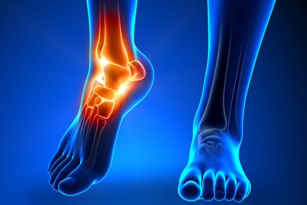
Calcaneal fractures: what they are, how to intervene
Fractures of the calcaneus (heel bone) often result from great force. The diagnosis is made by X-ray and, if necessary, CT scan. Treatment requires orthopaedic advice and includes plaster casts and sometimes surgery
Calcaneal fractures are serious but uncommon injuries
They account for only 1 and 2% of all fractures.
However, if not diagnosed and treated promptly, they can lead to long-term disability.
Up to 10% of these fractures are unrecognised at the initial visit to an emergency room.
Typically, these fractures are related to a high-energy trauma to the foot (e.g., a fall from a height onto the heels).
Because these fractures are caused by a large traumatic force, they are often accompanied by other serious injuries; 10% of patients with a calcaneal fracture have a thoracolumbar compression fracture.
Stress fractures can also occur in the calcaneus, especially in athletes, such as marathon runners.
Calcaneal fractures can be intra-articular.
Symptomatology of calcaneal fractures
Usually, the area around the heel and hindfoot is painful and very swollen.
Patients cannot put weight on their feet.
Acute compartment syndrome occurs in up to 10% of patients.
Diagnosis of calcaneal fractures
- X-RAY
- Sometimes CT scan
If a calcaneal fracture is suspected, X-ray examinations including axial and lateral projections should be performed.
CT scan is performed if
- X-rays are negative but clinical findings suggest a calcaneal fracture.
- Bohler’s angle is < 20°.
- More details regarding the fracture are required.

The Bohler angle is determined by lateral projection radiographs.
This angle is formed by the intersection of a line drawn from the superior apex of the posterior calcaneal tuberosity to the superior articular subastragalic surface and a line drawn from the superior articular subastragalic surface to the superior limit of the anterior calcaneal process.
Normally, the angle is 20-40°. An angle of < 20° indicates a fracture.
Physicians should also check for other injuries, such as thoracolumbar compression fractures and compartment syndrome.
Treatment of calcaneal fractures
Orthopaedic consultation
- Casting or possibly surgery, depending on the type of fracture
- Orthopaedic consultation is necessary.
There is much debate as to whether intra-articular calcaneal fractures should be treated surgically or non-surgically.
Extra-articular fractures of the calcaneus are treated symptomatically with protection, rest (avoiding loading), a compression bandage (including protection), ice and elevation (PRICE).
When the oedema resolves, a plaster is applied.
Read Also:
Emergency Live Even More…Live: Download The New Free App Of Your Newspaper For IOS And Android
Bone Cysts In Children, The First Sign May Be A ‘Pathological’ Fracture
Fracture Of The Wrist: How To Recognise And Treat It
Fractures Of The Growth Plate Or Epiphyseal Detachments: What They Are And How To Treat Them
Stress Fractures: Risk Factors And Symptoms