
Echo- and CT-guided biopsy: what it is and when it is needed
Echo- and CT-guided biopsy represents a system of tissue sampling for analysis. These take place with the aid of ultrasound or CT scans to see the target of the procedure while it is being performed
What are echo- and CT-guided biopsies used for?
Echo- and CT-guided biopsies involve the use of imaging techniques (ultrasound or CT scan) to insert a needle as precisely as possible into an organ.
The purpose of the manoeuvre is to take a tissue sample for subsequent histological analysis in order to obtain an accurate diagnosis and, in some cases, to prepare an appropriate therapy.
Their use avoids the need for a more invasive surgical procedure to take tissue for analysis.
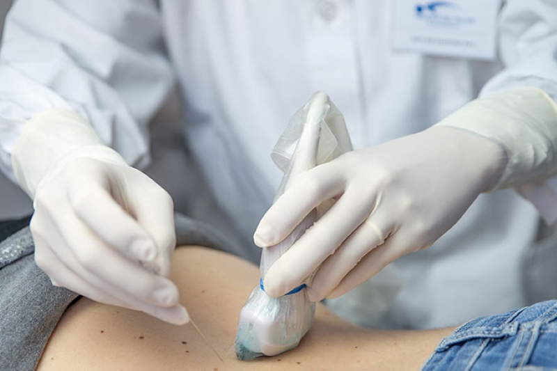
How does echo- and CT-guided biopsy work?
The manoeuvre usually takes a few minutes and involves local anaesthesia.
An agocannula is inserted into the patient’s arm and then the location where the needle is to be inserted is localised by ultrasound or CT scan.
The needle is then inserted to the point where the biopsy is to be taken, again guided by imaging.
The number of samples taken varies from case to case, a single sample being sufficient in the vast majority of cases.
Are preparation rules necessary?
The performance of echo- and CT-guided biopsies requires abstaining from food and drink for at least six hours prior to the procedure.
It may also be necessary to stop taking certain medications (especially anticoagulants and anti-platelets) in the 4-7 days preceding the biopsy.
On the day set for the procedure, it is necessary to come to the clinic with any tests that the doctor may require in anticipation of the biopsy.
Who can undergo an echo- and CT-guided biopsy?
The test may be contraindicated during pregnancy (if a CT scan is used). There may also be more specific contraindications that vary from case to case.
A liver biopsy, for example, cannot be performed if the patient suffers from coagulation disorders.
Is an echo- and CT-guided biopsy painful or dangerous?
The procedure is not painful.
The main risk is bleeding of the sampled tissue.
If a lung biopsy is performed, a secondary risk is the formation of a pneumothorax.

In this eventuality, the lung ‘deflates’ and it may be necessary to place a pleural drainage catheter to resolve the problem.
The use of ultrasound or CT scan minimises the risk of damage to organs and structures surrounding the target.
Ultrasonography is generally considered to be risk-free, while CT scans are associated with radiation risks, which are minimised by the reduction in radiation exposure.
Read Also
Emergency Live Even More…Live: Download The New Free App Of Your Newspaper For IOS And Android
Fusion Prostate Biopsy: How The Examination Is Performed
What Is Needle Aspiration (Or Needle Biopsy Or Biopsy)?
What Is Echocolordoppler Of The Supra-Aortic Trunks (Carotids)?
What Is The Loop Recorder? Discovering Home Telemetry
Cardiac Holter, The Characteristics Of The 24-Hour Electrocardiogram
Peripheral Arteriopathy: Symptoms And Diagnosis
Endocavitary Electrophysiological Study: What Does This Examination Consist Of?
Cardiac Catheterisation, What Is This Examination?
Echo Doppler: What It Is And What It Is For
Transesophageal Echocardiogram: What Does It Consist Of?
Venous Thrombosis: From Symptoms To New Drugs
Echotomography Of Carotid Axes