
Endocavitary electrophysiological study: what does this examination consist of?
The endocavitary electrophysiological study, also known by the acronym EES, is a diagnostic investigation aimed at examining the electrical properties of the heart in order to establish susceptibility to developing various heart rhythm disorders
Endocavitary electrophysiological study: what does it consist of?
This is an invasive examination, generally prescribed to investigate the nature and causes of different types of cardiac arrhythmias.
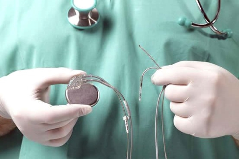
The EES is performed after a short hospitalisation and involves local anaesthesia; 2 to 5 catheters are inserted into the heart, which record the electrical signals originating from the heart cavities.
Generally, the main access routes are the right femoral vein at groin level, or the left subclavian vein at shoulder level; more rarely, it is possible to perform the examination via the jugular vein in the neck, or alternatively, in special cases, a trans-oesophageal electrophysiological study can be used.
In some cases provocative tests are performed, through the administration of drugs, whereby cardiac arrhythmia is induced and thus detected.
In this way, the cardiologist can study the electrocardiogram in detail, making it possible to locate the point or anatomical structure responsible for the disturbance.
Why an endocavitary electrophysiological study is performed
An endocavitary electrophysiological study is generally prescribed for patients at risk of developing cardiac arrhythmias for whom an arrhythmological diagnosis cannot be made using non-invasive methods: thanks to this study, it is possible to analyse the nature and mechanism of the arrhythmia in detail, enabling the cardiologist to make a more precise diagnosis of the type of cardiac disorder the patient is suffering from; identification of the arrhythmia also enables a suitable therapy to be developed for its resolution and/or prevention.
The purpose of the examination is to
- assess the functionality of the heart’s electrical conduction system
- identify the site and mechanism underlying the onset of arrhythmic phenomena;
- identify any paroxysmal symptoms (such as palpitations, syncope or lipotimia);
- assessing the effectiveness and side effects of certain drugs for arrhythmic prevention.
The EES is also generally used to treat cardiac arrhythmias that do not respond to pharmacological cardioversion therapy.
In particular:
- supraventricular tachycardia;
- Wolff-Parkinson-White syndrome;
- atrial fibrillation;
- atrial flutter;
- ectopic atrial tachycardia.
In most cases, during the procedure, the alterations in heart rhythm are interrupted using the same pulses that generated the arrhythmia.
In more severe and less responsive conditions, treatment of the arrhythmia involves one or more radiofrequency energy deliveries using a transcutaneous ablation procedure.
Like all invasive procedures, the endocavitary electrophysiological study has a minimal risk of complications such as local haematoma formation or injury to blood vessels and nerves.
Generally it is a well-tolerated examination: some patients may present with the same symptoms reported during the onset of arrhythmic episodes, such as palpitation, chest pain or dizziness; however, in most cases these resolve at the end of the procedure.
The average hospital stay is two nights, but after a few hours’ rest the patient is able to get up and resume normal activities.
How should I prepare for an endocavitary electrophysiological study
Before performing an endocavitary electrophysiological study, it is necessary to undergo a specialist cardiological evaluation, which includes the assessment of some laboratory tests, an electrocardiogram and an echocardiogram, to ensure maximum safety during the procedure.
This is in fact an invasive examination that requires a short hospitalisation and takes place in a specially equipped room.
Under local anaesthesia, 2 to 5 thin catheters are introduced through the veins in the groin or the clavicle region (subclavian vein), which are pushed into the heart cavities, where they record the electrical activity of the heart chambers.
The examination is practically painless and involves a small dressing at the site where the electro-catheters are introduced.
*This is indicative information: it is therefore necessary to contact the facility where the examination is performed to obtain specific information on the preparation procedure.
Read Also:
Head Up Tilt Test, How The Test That Investigates The Causes Of Vagal Syncope Works
What Is Ischaemic Heart Disease And Possible Treatments
Percutaneous Transluminal Coronary Angioplasty (PTCA): What Is It?
Ischaemic Heart Disease: What Is It?

EMS: Pediatric SVT (Supraventricular Tachycardia) Vs Sinus Tachycardia
Paediatric Toxicological Emergencies: Medical Intervention In Cases Of Paediatric Poisoning
Valvulopathies: Examining Heart Valve Problems
What Is The Difference Between Pacemaker And Subcutaneous Defibrillator?
Heart Disease: What Is Cardiomyopathy?
Inflammations Of The Heart: Myocarditis, Infective Endocarditis And Pericarditis
Heart Murmurs: What It Is And When To Be Concerned
Clinical Review: Acute Respiratory Distress Syndrome
Botallo’s Ductus Arteriosus: Interventional Therapy
Heart Valve Diseases: An Overview
Cardiomyopathies: Types, Diagnosis And Treatment
First Aid And Emergency Interventions: Syncope
Tilt Test: What Does This Test Consist Of?
Cardiac Syncope: What It Is, How It Is Diagnosed And Who It Affects
New Epilepsy Warning Device Could Save Thousands Of Lives
Understanding Seizures And Epilepsy
First Aid And Epilepsy: How To Recognise A Seizure And Help A Patient
Neurology, Difference Between Epilepsy And Syncope
Positive And Negative Lasègue Sign In Semeiotics
Wasserman’s Sign (Inverse Lasègue) Positive In Semeiotics
Positive And Negative Kernig’s Sign: Semeiotics In Meningitis
Lithotomy Position: What It Is, When It Is Used And What Advantages It Brings To Patient Care
Trendelenburg (Anti-Shock) Position: What It Is And When It Is Recommended
Prone, Supine, Lateral Decubitus: Meaning, Position And Injuries
Stretchers In The UK: Which Are The Most Used?
Does The Recovery Position In First Aid Actually Work?
Reverse Trendelenburg Position: What It Is And When It Is Recommended
Drug Therapy For Typical Arrhythmias In Emergency Patients
Canadian Syncope Risk Score – In Case Of Syncope, Patients Are Really In Danger Or Not?