
What is breast needle biopsy?
Needle biopsy is a breast tissue sampling technique used when there is diagnostic doubt regarding structural alterations revealed by previous diagnostic tests
Needle biopsy allows samples (tissue frustules) to be taken with a hollow needle through the skin for histological examination (histology is the study of the nature and characteristics of tissue).
This is a laboratory examination that allows accurate observation of the samples, ensuring with greater certainty than cytological examination (the examination of cells alone) the possibility of establishing whether the lesion is malignant or benign in nature (e.g. fibroadenoma).
What is breast needle biopsy used for?
The use of this technique is common in the field of oncology.
It is usually performed following mammograms and ultrasound scans that have revealed probable lesions or tumour formations or a cytological examination that has left some uncertainty about the final diagnosis.
It serves to ascertain through a laboratory study the nature of the tissue and possibly establish the pathology involved: a malignant or benign tumour.
Breast needle biopsy, are there any preparation rules?
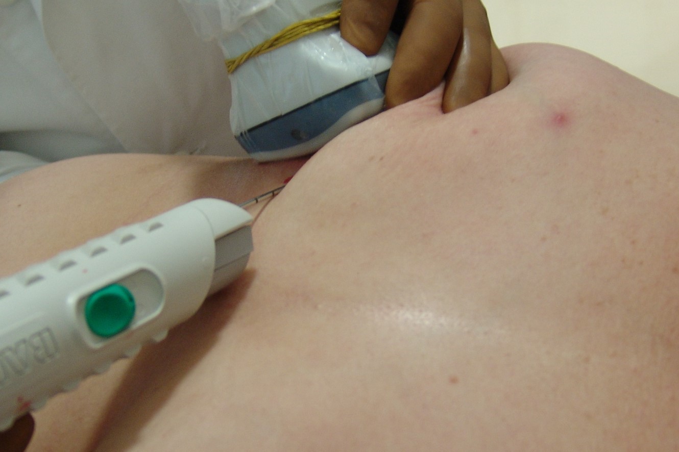
Needle biopsy consists of targeted sampling with a hollow needle that is introduced through the skin at the site of the lesion.
Generally, this operation is performed under ultrasound guidance.
The operator identifies the precise target of the sampling thanks to real-time observation on a screen of the images processed by the ultrasound scanner.
The rules of preparation for the person undergoing the procedure are the suspension of any therapy with anticoagulants on the advice of the referring doctor or TAO centre or senologist.
The person must wear comfortable clothes.
It will be the caregiver’s responsibility to prepare her so that the area is free and accessible for the type of collection.
The patient must be accompanied, because even if hospitalisation is not planned, the effects of anaesthesia and any temporary discomfort do not allow her to return to her home alone.
Who can perform the examination?
Needle biopsy, unlike needle aspiration, is more invasive.
This means that the needle used and the type of sample have a greater effect on the person.
It is performed under local anaesthesia, through a small incision in the indicated area.
It therefore presents the normal precautions associated with the use of analgesic drugs, agreed upon with the specialist, but the frequent and common use of this method has no contraindications, except in very rare cases.
How does breast needle biopsy work?
A needle biopsy is performed under local anaesthesia and a small incision must be made in the skin.
The person is made to lie supine on a couch with their arms overhead and the breast area uncovered.
Only in some cases may the patient be asked to lie on her side.

Immediately after the sampling it may be helpful to manually compress the affected area for a few minutes to reduce the risk of bleeding.
The examination lasts between 20 and 30 minutes and at the end a bag of dry ice is applied to accelerate the absorption of any bruising and relieve the painful sensation.
The person can return home immediately afterwards and a rest period of a few hours is recommended.
In the following days, discomfort may occur in the area of the puncture, while the presence of small bruise-like patches coinciding with the small amount of blood in the tissues surrounding the spot should not be frightening.
Read Also
Emergency Live Even More…Live: Download The New Free App Of Your Newspaper For IOS And Android
Fusion Prostate Biopsy: How The Examination Is Performed
CT (Computed Axial Tomography): What It Is Used For
What Is An ECG And When To Do An Electrocardiogram
MRI, Magnetic Resonance Imaging Of The Heart: What Is It And Why Is It Important?
Mammary MRI: What It Is And When It Is Done
What Is Needle Aspiration (Or Needle Biopsy Or Biopsy)?
Positron Emission Tomography (PET): What It Is, How It Works And What It Is Used For
CT, MRI And PET Scans: What Are They For?
MRI, Magnetic Resonance Imaging Of The Heart: What Is It And Why Is It Important?
Urethrocistoscopy: What It Is And How Transurethral Cystoscopy Is Performed
What Is Echocolordoppler Of The Supra-Aortic Trunks (Carotids)?
Surgery: Neuronavigation And Monitoring Of Brain Function
Robotic Surgery: Benefits And Risks
Refractive Surgery: What Is It For, How Is It Performed And What To Do?
Single Photon Emission Computed Tomography (SPECT): What It Is And When To Perform It