
Hypoxemia: meaning, values, symptoms, consequences, risks, treatment
The term ‘hypoxaemia’ refers to an abnormal decrease in oxygen content in the blood, caused by an alteration in gas exchanges occurring in the pulmonary alveoli
About hypoxaemia: normal and pathological values
Hypoxemia occurs when the partial pressure of oxygen in arterial blood (PaO2) is less than 55-60 mmHg and/or the oxygen saturation of haemoglobin (SpO2) is less than 90%.
Recall that oxygen saturation normally ranges between 97% and 99% in healthy subjects, while it may be physiologically lower in the elderly (around 95%) and severely lower (at or below 90%) in subjects with pulmonary and/or circulatory diseases.
If PCO2 is above 45 mmHg at the same time, hypoxaemia occurs along with hypercapnia, i.e. an abnormal increase in the concentration of carbon dioxide (CO2) in the blood.
Normal PaO2 values vary greatly according to age (higher in the young, lower in the elderly), but are normally between about 70 and 100 mmHg: a PaO2 below 70 mmHg reveals mild hypoxia, while when it falls below 40 mmHg it indicates particularly severe hypoxemia.
Causes
Hypoxaemia is caused by an abnormal and more or less severe decrease in gas exchange between blood and atmosphere that occurs in the pulmonary alveoli; this alteration occurs for various causes, acute and chronic.
Acute hypoxaemia causes
- asthma;
- pulmonary oedema;
- pneumonia;
- pneumothorax
- respiratory distress syndrome (ARDS);
- pulmonary embolism;
- mountain sickness (above an altitude of 2,500 metres);
- drugs that depress the activity of respiratory centres, e.g. narcotics (such as morphine) and anaesthetics (such as propofol).
Causes of chronic hypoxaemia:
- emphysema;
- pulmonary fibrosis;
- chronic obstructive pulmonary disease (COPD);
- pulmonary neoplasms;
- interstitial lung diseases;
- congenital heart defects;
- brain lesions.
Symptoms and signs
Hypoxaemia itself is a sign of a disease or condition; depending on the cause, hypoxaemia may be associated with various symptoms and signs, including:
- cyanosis (bluish skin);
- cherry-red coloured skin;
- general malaise;
- dyspnoea (difficulty breathing);
- Cheyne-Stokes respiration;
- apnoea;
- arterial hypertension;
- arrhythmias;
- tachycardia;
- ventricular fibrillation;
- cardiac arrest;
- confusion;
- coughing;
- haemoptysis (emission of blood from the respiratory tract);
- tachypnoea (increased respiratory rate);
- sweating;
- asthenia (lack of strength);
- hippocratic (drumstick) fingers;
- low oxygen saturation;
- low partial pressure of oxygen in the blood.
- coma and death in the most severe cases.
Not all the symptoms listed are always present at the same time.
In the case of simultaneous hypercapnia, one may also experience:
- reddening of the skin;
- elevated heart rate;
- extrasystoles;
- muscle spasms
- reduced brain activity
- increased blood pressure;
- increased cerebral blood flow;
- headaches;
- confusion and lethargy;
- increased cardiac output.
In the case of severe hypercapnia (PaCO2 generally exceeding 75 mmHg), the symptoms progress to disorientation, panic, hyperventilation, convulsions, loss of consciousness, and may even lead to death.
Remember, however, that hypoxaemia is on average more severe and more rapidly fatal than hypercapnia.
Consequences
The possible consequence of hypoxemia is hypoxia, i.e. a decrease in the amount of oxygen available in the tissue, which can lead to necrosis (i.e. death) of the tissue where it occurs, since oxygen is necessary for cell survival.
Hypoxia can be ‘generalised’ (i.e. affecting the entire organism) or ’tissue-based’ when oxygen deficiency affects a specific tissue of the organism (e.g. the dreaded cerebral hypoxia, which can lead to irreparable damage and even death in the most severe cases).
Diagnosis
Diagnosis is based on anamnesis, objective examination and a number of possible laboratory and imaging tests (such as chest X-ray or endoscopy).

Two basic parameters for establishing the state of hypoxemia are:
- oxygen saturation (SpO2): measured with a saturation meter (a kind of clothes peg that is applied for a few seconds on a finger, non-invasively);
- partial pressure of oxygen in arterial blood (PaO2): measured with haemogasanalysis, a more invasive test in which blood is taken from the patient’s wrist with a syringe.
Depending on the patient’s age and PaO2 mmHg, hypoxia is classified as mild, moderate or severe:
- mild hypoxia: PaO2 of approximately 60 – 70 mmHg (below 80 mmHg if the patient is under 30 years of age);
- moderate hypoxia: PaO2 40 – 60 mmHg;
- severe hypoxia: PaO2 < 40 mmHg.
SpO2 values correlate with PaO2 values: an SpO2 value of 90% generally correlates with a PaO2 value of less than 60 mmHg.
Therapy
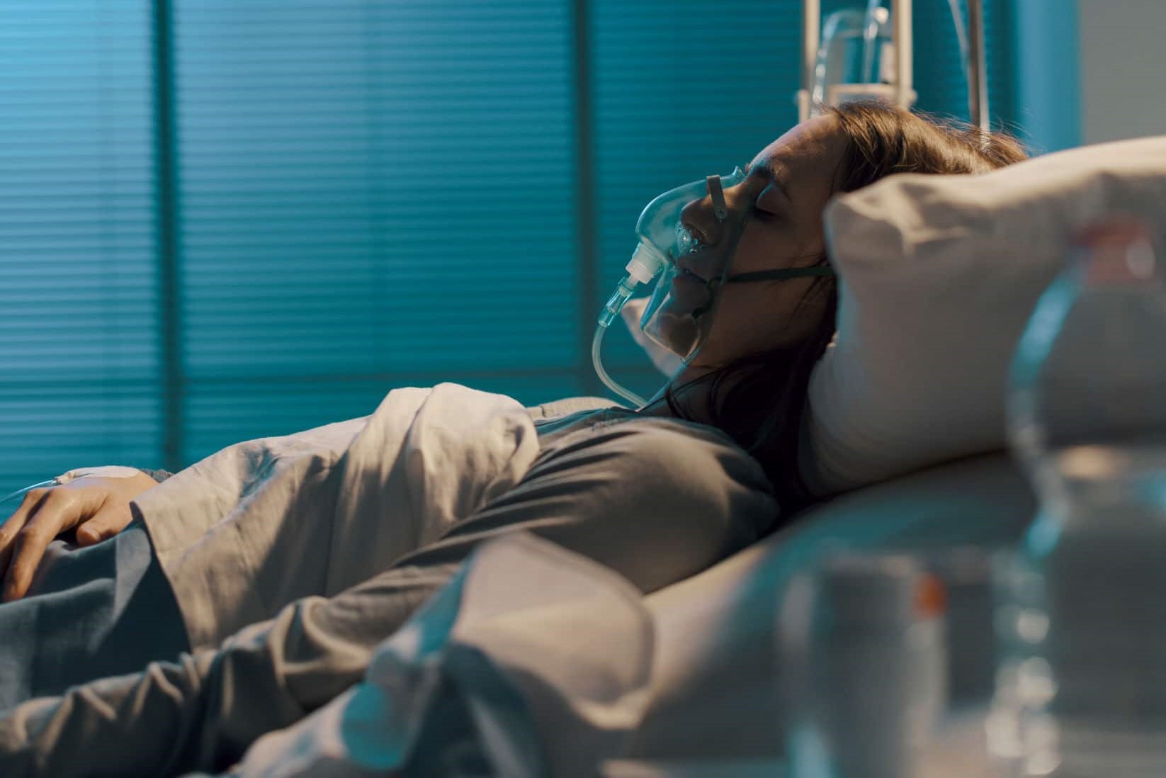
The hypoxaemic patient must first be treated with oxygen administration (oxygen therapy) and, in severe cases, with assisted ventilation.
Secondly, the underlying cause must be determined and this cause specifically treated, e.g. in the case of severe asthma, the patient must be given bronchodilators or inhaled corticosteroids.
Read Also
Emergency Live Even More…Live: Download The New Free App Of Your Newspaper For IOS And Android
Obstructive Sleep Apnoea: What It Is And How To Treat It
Difference Between Hypoxaemia, Hypoxia, Anoxia And Anoxia
Occupational Diseases: Sick Building Syndrome, Air Conditioning Lung, Dehumidifier Fever
Obstructive Sleep Apnoea: Symptoms And Treatment For Obstructive Sleep Apnoea
Our respiratory system: a virtual tour inside our body
Tracheostomy during intubation in COVID-19 patients: a survey on current clinical practice
FDA approves Recarbio to treat hospital-acquired and ventilator-associated bacterial pneumonia
Clinical Review: Acute Respiratory Distress Syndrome
Stress And Distress During Pregnancy: How To Protect Both Mother And Child
Respiratory Distress: What Are The Signs Of Respiratory Distress In Newborns?
Pneumology: Difference Between Type 1 And Type 2 Respiratory Failure