
Pathophysiology of thoracic trauma: injuries to the heart, great vessels and diaphragm
Injuries of the heart as a consequence of thoracic trauma: trauma is currently one of the most serious public health problems worldwide
In industrialised countries, they are the leading cause of death in the under-40 age group and the third leading cause of death after heart disease and cancer.
In about a quarter of cases, injuries lead to a disability that requires the patient to be bedridden and undergo complex treatment and rehabilitation.
Given the young age of most of these patients, trauma is responsible – economically speaking – for more severe disability and loss of productivity overall than even heart disease and cancer taken together.
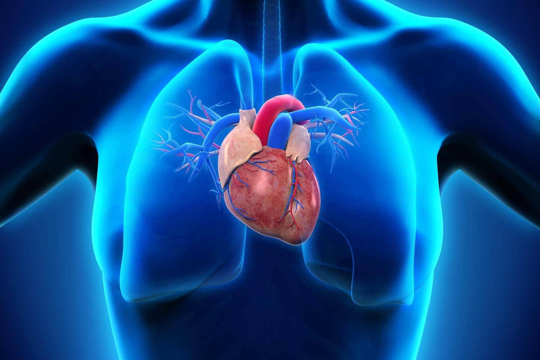
Pathophysiology of thoracic trauma: injuries to the heart and great vessels
Trauma to the chest can cause various forms of cardiac injury, such as penetration of a foreign body, rupture, tamponade, laceration and occlusion of the coronary arteries, myocardial contusion, pericardial effusion, septal defects, valvular lesions, rupture of the great vessels.
These injuries are often rapidly fatal.
Penetrating cardiac injuries are most often caused by blunt weapons or shotguns and result in a mortality rate of between 50% and 85%.
Closed traumas are more frequently associated with a rupture of the heart (the right ventricle is affected more often than the left) and result in a mortality rate of about 50 per cent in patients who arrive in the emergency room alive.
After a rupture of a heart chamber or a tear in the coronaries or large vessels, blood rapidly fills the pericardial sac and results in cardiac tamponade.
Even as little as 60-100 ml of blood can cause cardiac tamponade and cardiogenic shock, resulting from a reduction in diastolic filling.
Puncture wounds penetrating the pericardial sac and inside the heart result in rapid haemorrhage, which dominates the clinical picture.
Interestingly, cardiac tamponade following a gunshot wound to the heart is associated with increased survival due to systemic hypotension and increased pressure in the pericardial space, which help to limit haemorrhage.
A cardiac tamponade is often associated with the clinical symptoms of Beck’s triad (jugular venous distension, hypotension and attenuation of cardiac tones).
This triad, however, may not be present in patients who have become hypovolaemic due to haemorrhage. radiographic evidence of a widening of the mediastinal shadow may suggest an effusion in the mediastinum and/or tamponade.
Confirmation of a pericardial effusion can be more easily and accurately provided by echocardiography.
The therapeutic measures of choice consist of an emergency exploratory thoracotomy, with cardiopulmonary bypass and surgical correction, and transfusion as required by the clinical condition.
It is not easy to identify a myocardial contusion after closed chest trauma but, in carefully monitored patients, the incidence is probably close to 25%.
The anatomopathological changes of the contused heart consist of intramyocardial haemorrhages, myocardial oedema, coronary occlusion, myofibrillar degeneration and necrosis of myocardiocytes.
These lesions lead to arrhythmias and haemodynamic instability similar to those observed after a myocardial infarction.
On the electrocardiogram (ECG), tachycardia, ST-segment elevation, T-wave changes and occasional ventricular premature contractions are often present (3,25,29).
Plasma enzymes (glutamic oxalacetic transaminase [GOT], lactate dehydrogenase [LDH] and creatine phosphokinase [CPK]) are almost always elevated after closed chest trauma and are therefore of little diagnostic value.
An increase in the CPK-MB isoenzyme appears to have greater discriminating power and contributes to the diagnosis of myocardial contusion.
Pulmonary artery catheterisation is often useful for monitoring haemodynamic conditions and for the treatment of possible decompensation.
The battery of examinations for the identification of a myocardial contusion includes:
- echocardiogram,
- radionuclide angiography,
- serial electrocardiographic examinations,
- determination of haemodynamic parameters,
- monitoring of CPK-MB levels.
Treatment is the same as for myocardial infarction.
In patients with heart failure, the application of an aortic counterpulsator has proven useful in improving cardiac output.
Often there is complete healing, which leaves only minimal scarring at the myocardial level.
The overall mortality rate of patients with myocardial contusion is about 10%
Rupture of the aorta, caused by closed chest trauma (e.g. in a car accident) and subsequent exsanguination lead to a dramatic situation and rapidly result in the death of the patient, often without the physician being able to intervene in time.
In the United States, approximately 8-10 thousand people a year suffer a ruptured aorta and, of these, approximately 80-90% die within minutes.
In patients who still arrive at the hospital alive, the injury is to the proximal portion of the descending thoracic aorta.
Patients usually present severely hypotensive and often with radiographic signs of mediastinal enlargement.
The diagnostic method of choice when suspecting an aortic rupture or tear is aortography.

In the presence of shock or obvious mediastinal widening, an emergency thoracotomy is necessary, with surgical correction of the lesion, with transfusions as required by the patient’s clinical condition.
Pathophysiology of thoracic trauma: diaphragmatic injuries
The most frequent cause of diaphragmatic injuries is penetrating trauma.
Closed abdominal trauma results in a rupture of the diaphragm in only 5% of cases.
A rupture of the diaphragm is associated with rupture of the spleen, haemothorax, reduced mobility of the diaphragm itself, shock, ventilatory failure, retention of CO2, coma, intestinal herniation in the thorax, resulting in intestinal stricture and reduced lung volume.
Mortality in this clinical setting has been estimated at 29%, but certainly such a high rate is related to other associated injuries, rather than diaphragmatic involvement alone.
Diagnosis is usually made on the basis of the results of a chest and abdominal X-ray, CT scan or during an exploratory laparotomy. Rupture of the diaphragm requires surgical evaluation and correction.
Contusion and weakening of the diaphragm are diagnosed much less frequently and are probably associated with difficult ventilation and a reduction in the patient’s ability to cough.
Heart injuries: late complications of chest wall trauma
Chronic pain, recurrent atelectasis and pneumonia are the most frequent late and protracted complications of chest trauma.
In most cases, their cause remains undetermined and treatment consists of reassuring patients and administering analgesics.
Occasionally, surgery is required to correct rib or sternal fractures that are responsible for persistent pain symptoms.
A pleural infection may be due to an undrained haemothorax or the retention of a foreign body and may evolve into pleurisy, empyema or fibrothorax.
Thoracotomy, pleural drainage, administration of antibiotics, and decortication of the pleura are all treatments frequently practised in cases of pleural infections that are unresponsive to other therapies, to prevent the formation of a fibrothorax.
Both closed and penetrating trauma can lead to the appearance of an arteriovenous fistula, aortic aneurysm, cardiac valve insufficiency, or constrictive pericarditis, diaphragmatic herniation, stenosis or oesophageal fistulas.
A retained foreign body may migrate or penetrate into different regions, even many years later.
Migration of a foreign body can also lead to embolic events. Tissue erosion by a sharp foreign body may be responsible for haemoptysis, pneumonia or lung abscesses.
The treatment of these long-term complications often requires surgical correction along with care during the acute phase and a period of rehabilitation.
Read Also:
Emergency Live Even More…Live: Download The New Free App Of Your Newspaper For IOS And Android
Recovery For Broken Ribs: What To Do, How Long It Takes
Tracheal Intubation: When, How And Why To Create An Artificial Airway For The Patient
What Is Transient Tachypnoea Of The Newborn, Or Neonatal Wet Lung Syndrome?
Traumatic Pneumothorax: Symptoms, Diagnosis And Treatment
Diagnosis Of Tension Pneumothorax In The Field: Suction Or Blowing?
Pneumothorax And Pneumomediastinum: Rescuing The Patient With Pulmonary Barotrauma
ABC, ABCD And ABCDE Rule In Emergency Medicine: What The Rescuer Must Do
Multiple Rib Fracture, Flail Chest (Rib Volet) And Pneumothorax: An Overview
Internal Haemorrhage: Definition, Causes, Symptoms, Diagnosis, Severity, Treatment
Cervical Collar In Trauma Patients In Emergency Medicine: When To Use It, Why It Is Important
KED Extrication Device For Trauma Extraction: What It Is And How To Use It
How Is Triage Carried Out In The Emergency Department? The START And CESIRA Methods
Chest Trauma: Clinical Aspects, Therapy, Airway And Ventilatory Assistance
Pain Management In Blunt Thoracic Trauma