
Silicosis: causes, types, symptoms, diagnosis, pathological anatomy, treatment
Silicosis is a type of pneumoconiosis, i.e. a lung disease caused by the inhalation of dust, e.g. at work, and in fact silicosis is a typical occupational respiratory disease
Silicosis is usually caused by inhaling dust of free crystals of silicon (silicon dioxide, quartz) and is characterised by pulmonary fibrosis with distinct nodules and, in more advanced stages, by fibrosis with confluent nodules and impaired respiratory function.
Silicosis is undoubtedly the longest known occupational respiratory disease.
Causes of silicosis
Silicosis generally arises after prolonged inhalation of small particles of free crystalline silica in metal mines (lead, anthracite, copper, silver, gold), foundries, ceramic factories and sandstone and granite mining.
Free silica crystals are one form of free silica, a relatively pure form of silica not combined with silicidic acid.
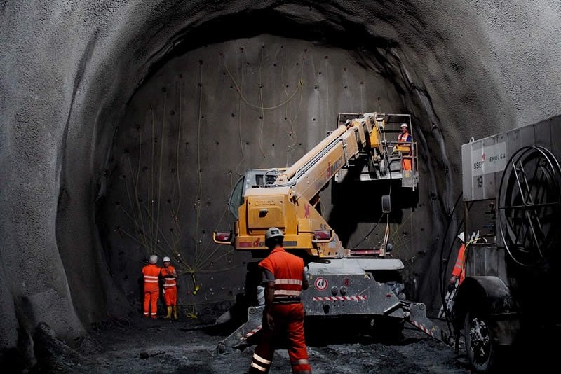
It usually takes 20-30 years of exposure before the disease becomes manifest, although it develops in < 10 years when exposure to dust is very high, such as in tunnel construction, abrasive soap factories and during blasting operations.
The current limit for free silica in the air of industrial environments is 100 mg/m3, an 8-hour time-weighted average based on the percentage of silica in the dust.
The formula for ascertaining whether the limit is exceeded is mg/m3.
Who is at risk?
Workers who inhale dust containing more than 1% are most exposed, particularly high-risk occupations are:
- working in mines;
- stone cutting;
- production of abrasives;
- foundry work;
- glass or ceramic production work;
- work in the refractory industry;
- surface cleaning or the discolouration of jeans by sandblasting.
However, the hazard depends not only on the type of work, but also on the percentage of crystalline silicon dioxide in the inhaled air and the length of time it is in the environment.
Pathological anatomy and pathophysiology of silicosis
Respirable free silica particles are phagocytosed by alveolar macrophages and enter the lymphatics and interstitial tissue.
The macrophages cause the release of cytotoxic enzymes that induce fibrosis of the lung parenchyma.
When a macrophage dies, silica particles are released and phagocytosed by other macrophages and the process may repeat.
The typical initial lesion is the formation of distinct hyaline silica nodules in both lungs.
The dying macrophages release silica into the interstitial tissue around the second generation of respiratory bronchioles, where a nodule forms.
Subsequently, fusion of the fibrotic nodules results in confluent masses, with retraction of the upper portions of the lung and emphysema and marked distortion of the lung architecture.
Ventilation and gas exchange are impaired.
The reduction of all lung volumes distinguishes the overall pathophysiological picture of confluent silicosis from that of advanced pulmonary emphysema.
In the advanced stages of confluent silicosis, severe functional impairments occur and respiratory failure, their extreme consequence, may progress along with a worsening of the rx picture for a limited time (i.e., 2-5 years) even after exposure has stopped.
When dust exposure is extremely high and silico-proteinosis develops, the alveolar spaces fill with a proteinaceous material similar to that found in alveolar proteinosis and mononuclear cells infiltrate the septa.
Symptoms and signs
Patients with simple nodular silicosis have neither symptoms nor, usually, respiratory impairment.
They may complain of coughing and expectoration, but these symptoms are due to industrial bronchitis and occur with the same frequency in subjects with normal rx.
Although uncomplicated silicosis impairs respiratory function to a small extent, patients with stage 2 or 3 disease (see diagnosis, below) occasionally have a slight reduction in lung volumes, but values rarely fall below reference limits.
Confluent nodule silicosis, on the other hand, may result in severe dyspnoea, coughing and sputum
The severity of breathlessness is related to the size of the confluent masses in the lungs.
When the masses are very large, the patient achieves severe disability.
As the masses invade and obliterate the vascular bed, pulmonary hypertension and right ventricular hypertrophy set in.
In advanced stages, objective signs of consolidation and pulmonary hypertension may be present.
Non-hypoxaemic pulmonary heart disease is ultimately a cause of death.
In confluent (complicated) silicosis, especially in the terminal stages, alterations in respiratory function are common.
These include decreased lung volumes and diffusing capacity and airway obstruction, often with pulmonary hypertension and occasionally mild hypoxaemia.
CO2 retention is rare.
In many silica patients, the serum contains anti-polon autoantibodies and antinuclear factors.
Individuals exposed to silica in the work environment and who have a positive tuberculin test have an increased risk of developing TB.
In general, the greater the amount of silica present in the lungs, the higher the risk.
Diagnosis
Diagnosis is based on characteristic x-ray changes and a history of exposure to free silica.
Simple silicosis is recognised by the presence of numerous small round or regular opacities on chest X-ray and is classified into categories 1, 2 and 3 according to their prevalence.
Confluent nodule silicosis is revealed by the presence of an opacity > 1 cm in diameter on a background of category 2 or 3 simple silicosis.
A number of other diseases that may mimic simple silicosis include milar TB, welders’ siderosis, sarcoidosis, haemosiderosis and coal miners’ pneumoconiosis.
However, the presence of ‘eggshell’ calcifications in the hilar and mediastinal lymph nodes distinguishes silicosis from other occupational respiratory diseases.
Silico-tuberculosis resembles confluent nodule silicosis on X-ray.
Differentiation can be made by culture test of sputum.
Prophylaxis and therapy for silicosis
Effective dust control can prevent silicosis.
Since dust reduction systems cannot reduce the risk in sandblasting workers, masks supplied with outside air must be worn.

However, these measures may not be available for personnel in the area who are engaged in other activities (e.g. painters, welders).
It is therefore preferable to replace sand with other abrasive materials.
Monitoring of all exposed workers is by periodic chest x-rays q 6 months for sandblasters and q 2-5 years for other exposed workers.
No other effective treatment other than lung transplantation is known.
Individuals with airway obstruction must be treated as for chronic airway obstruction.
Those exposed to silica who have a positive tuberculin reaction but a negative sputum culture test for TB should take isoniazid for at least 1 year.
Some experts recommend lifelong treatment because the function of alveolar macrophages may be permanently impaired by silica.
Lifetime prophylaxis with isoniazid may be indicated for those who have been previously treated for active TB.
Patients with silicosis and active pulmonary TB require an extension of standard multidrug therapy by at least 3-6 months.
Other occupational respiratory diseases
Other frequent occupational respiratory diseases that might interest you are:
- coal workers’ pneumoconiosis
- asbestosis and related diseases (mesothelioma and pleural effusion);
- berylliosis;
- hypersensitivity pneumonias;
- occupational asthma;
- byssinosis;
- diseases caused by irritant gases and other chemicals;
- sick building syndrome.
Read Also
Emergency Live Even More…Live: Download The New Free App Of Your Newspaper For IOS And Android
Symptoms Of Asthma Attack And First Aid To Sufferers
Occupational Asthma: Causes, Symptoms, Diagnosis And Treatment
Bronchial Asthma: Symptoms And Treatment
Bronchitis: Symptoms And Treatment
Bronchiolitis: Symptoms, Diagnosis, Treatment
Extrinsic, Intrinsic, Occupational, Stable Bronchial Asthma: Causes, Symptoms, Treatment
Chest Pain In Children: How To Assess It, What Causes It
Bronchoscopy: Ambu Set New Standards For Single-Use Endoscope
What Is Chronic Obstructive Pulmonary Disease (COPD)?
Respiratory Syncytial Virus (RSV): How We Protect Our Children
Respiratory Syncytial Virus (RSV), 5 Tips For Parents
Infants’ Syncytial Virus, Italian Paediatricians: ‘Gone With Covid, But It Will Come Back’
Respiratory Syncytial Virus: A Potential Role For Ibuprofen In Older Adults’ Immunity To RSV
Neonatal Respiratory Distress: Factors To Take Into Account
Stress And Distress During Pregnancy: How To Protect Both Mother And Child
Respiratory Distress: What Are The Signs Of Respiratory Distress In Newborns?
Respiratory Distress Syndrome (ARDS): Therapy, Mechanical Ventilation, Monitoring
Bronchiolitis: Symptoms, Diagnosis, Treatment
Chest Pain In Children: How To Assess It, What Causes It
Bronchoscopy: Ambu Set New Standards For Single-Use Endoscope
Bronchiolitis In Paediatric Age: The Respiratory Syncytial Virus (VRS)
Pulmonary Emphysema: Causes, Symptoms, Diagnosis, Tests, Treatment
Bronchiolitis In Infants: Symptoms
Fluids And Electrolytes, Acid-Base Balance: An Overview
Ventilatory Failure (Hypercapnia): Causes, Symptoms, Diagnosis, Treatment
What Is Hypercapnia And How Does It Affect Patient Intervention?
Colour Changes In The Urine: When To Consult A Doctor
The Colour Of Pee: What Does Urine Tell Us About Our Health?
First Aid For Dehydration: Knowing How To Respond To A Situation Not Necessarily Related To The Heat
How To Choose And Use A Pulse Oximeter?
Alterations In Acid-Base Balance: Respiratory And Metabolic Acidosis And Alkalosis