
Horton's arteritis: symptoms, diagnosis and treatment of this vasculitis
Also called giant cell arteritis, gigantocellular arteritis or temporal arteritis, Horton’s arteritis is a form of vasculitis that mainly affects the arteries of the head and neck
A possible cause of headaches among the over-50s, it is much more common among women than men.
And, according to estimates, it affects 1-5 people per 10,000.
Characterised by headache and pain in the scalp and jaw, in severe cases it can generate fever, malaise and necrosis of the tissues supplied by the end of the arteries.
The causes are not yet known, but recent studies have shown that a combination of environmental and genetic factors underlie it.
Horton’s arteritis is an inflammation of the blood vessels especially affecting the arteries of the head and neck region
Most prevalent in the adult female population, it seems to affect Northern European populations most.
It is usually diagnosed between the ages of 70 and 75 as, especially in its early stages, its symptoms are not so distinguishable.
Instead, the symptomatology is very similar to that of a headache, with headaches, scalp hypersensitivity, jaw claudication and loss of vision.
Its most serious complication is precisely ocular symptoms, found in 20-30% of patients and sometimes responsible for irreversible monocular blindness.
Arteries are in fact flexible: their walls, although thick, are elastic.
The function they perform is fundamental.
They serve to transport oxygen-rich blood from the left ventricle of the heart to the organs and tissues, via the aorta, smaller and smaller arteries and up to the capillaries.
When arteries become inflamed, they enlarge abnormally and obstruct normal blood flow.
It is at this point that the symptoms of Horton’s arteritis originate.
Horton’s arteritis has no known cause to date
Scholars agree in attributing a combination of genetic and environmental factors (bacterial or viral infections) as possible causes for its onset.
However, there are certain risk factors, starting with gender and age.
The disease very rarely strikes under the age of 50-55, and becomes significantly more frequent from 65-70 onwards.
Women are affected twice as often as men, and the highest number of cases is recorded in Northern Europe (especially in the Scandinavian peninsula).
The main risk factor, however, is polymyalgia rheumatica: 15% of sufferers also suffer from Horton’s arteritis.
An inflammation of the muscles, polymyalgia rheumatica initially originates in the shoulder and neck muscles and then spreads throughout the body.
Also caused by a combination of genetic and environmental factors, it causes pain and muscle stiffness.
Early diagnosis of Horton’s arteritis is not easy
In its early stages it can easily be mistaken for a normal flu but, unlike the latter, it causes a headache – localised at the temples – that does not go away even with common antipyretics.
As the days go by, the symptomatology worsens, and the headache is accompanied by a sense of soreness in the temporal area of the skull, jaw pain and vision problems (double vision or loss of vision).
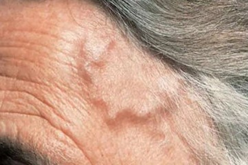
The typical symptom is therefore pain in the temples (usually bilateral but also unilateral or on the forehead).
But the patient may also experience:
- pain in the scalp, when resting the head or combing the hair
- fever, tiredness, pain and stiffness in the shoulders, neck and joints
- pain in the jaw
- weight loss without apparent cause
- swelling of the affected arteries
- pain in the tongue, especially when speaking and eating
Paying attention to these symptoms is essential as, if neglected, Horton’s arteritis can lead to serious complications
The most frequent is blindness: the blood vessels swell and narrow, and the eyes are less well supplied with blood.
Vision is thus impaired and a progressive loss arises which, if left untreated, can lead to total blindness.
Other, rarer complications of the disease are aortic aneurysm and stroke.
The former is a permanent abnormal dilatation of a small section of the aorta (if the aorta ruptures, the patient may suffer a fatal internal haemorrhage); the latter is caused by obstruction of the arterial vessels by a blood clot.
The diagnosis of Horton’s arteritis occurs in several stages
The examination begins with an objective test.
The doctor inquires about the patient’s medical and family history, verifies that he or she is not suffering from polymyalgia rheumatica, notes his or her symptoms and checks that the temporal arteries are not swollen and hard. If he suspects arteritis, he will prescribe specific blood tests: the ESR (erythrocyte sedimentation test) evaluates the speed at which red blood cells are deposited at the bottom of the tube.

The faster they settle, the higher the ESR, the more likely it is that inflammation is present.
Another important indicator is the C-reactive protein, produced by the liver: high values are a sign of an inflammatory state.
If necessary, it is possible to undergo instrumental tests to be certain of the diagnosis (but also at a later stage, to assess whether the therapy is effective):
- magnetic resonance imaging with contrast medium makes it possible to see how the inflammation has altered the blood vessels;
- the ecodoppler ‘photographs’ arteries and veins;
- the PET scan injects a radiopharmaceutical into the arteries to assess that the arteries have not changed due to inflammation (unlike the other tests, this one is more invasive as ionising radiation is used).
The most useful test for the diagnosis of Horton’s arteritis, however, is the biopsy
With the patient under local anaesthesia, the doctor removes a small piece of the temporal artery to observe it under a microscope.
If the cells are larger than they should be (but care must be taken to take genuinely inflamed portions of the artery), the diagnosis is certain.
Therapies
Horton’s arteritis is treated with corticosteroids because of their strong anti-inflammatory capacity.
However, this is a lengthy process.
If the first effects are seen after a few days, it takes 12-24 months to recover from Horton’s arteritis.
It is therefore necessary to continue treatment for a long time, periodically checking ESR and C-reactive protein levels: when their values return to normal, the patient can be said to be cured.
Especially in older patients, however, the erythrocyte sedimentation rate cannot be the only parameter considered.
Corticosteroids, although after the first moment they are slowly reduced until the minimum dose that produces an effect is found, are strong drugs that are not without side effects.
Their intake is correlated with an increased risk of developing osteoporosis, high blood pressure, diabetes, muscle weakness, glaucoma, cataracts, weight gain, weakened immune system and skin hypersensitivity.
Precisely because of the side effects, taking corticosteroids requires a certain amount of caution.
The patient must modify his diet to avoid the onset of hypertension and hyperglycaemia, eliminating sweets and fats and favouring fruit, vegetables and low-fat foods.
He should then exercise, and take vitamin D and calcium supplements.
Currently, however, there is a tendency to reserve corticosteroids for the most serious cases, those in which early intervention is necessary to prevent loss of sight.
Recently, the AIFA (Italian Medicines Agency) approved the use of tocilizumab as the first drug against Horton’s arteritis.
Administered subcutaneously, in combination with prednisone (thus used in lower doses), it resulted in a complete remission of the disease without symptoms in the patients tested.
Already used for rheumatoid arthritis in moderate and severe cases, systemic juvenile idiopathic arthritis and polyarticular juvenile idiopathic arthritis, it is now also used for giant cell arteritis (but not intravenously).
All patients, if there are no contraindications, must then be treated with low-dose aspirin to prevent ischaemic events.
Read Also
Emergency Live Even More…Live: Download The New Free App Of Your Newspaper For IOS And Android
Vasculitis: Symptoms And Causes Of Horton’s Arteritis
Venous Thrombosis: From Symptoms To New Drugs
Deep Vein Thrombosis Of The Upper Limbs: How To Deal With A Patient With Paget-Schroetter Syndrome
Venous Thrombosis: What It Is, How To Treat It And How To Prevent It
Non-Traumatic Intramural Hematomas In Patients On Anticoagulant Therapy
The New Oral Anticoagulants: Benefits, Dosages And Contraindications
Non-Traumatic Intramural Hematomas In Patients On Anticoagulant Therapy
Thrombus: Causes, Classification, Venous, Arterial And Systemic Thrombosis
Anticoagulant Drugs: List And Side Effects
Virchow’s Triad: The Three Risk Factors For Thrombosis