
Nevi: what they are and how to recognise melanocytic moles
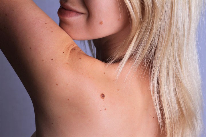
Nevi more commonly called melanocytic moles (or nevocellular nevi) are intracutaneous collections of melanocytes that clinically manifest as blotchy or nodular lesions with clear-cut borders and relatively stable over time
Nevus cells may be located intraepidermally (junctional nevus), dermo-epidermally (compound nevus) or intradermally (dermal nevus).
How to recognise nevi
In most cases nevi are asymptomatic and only rarely are they felt as a ‘presence’ or noticed because of their size or location.
Their clinical appearance is polymorphous: several varieties can be distinguished.
Flat melanocytic nevus: hyperchromic spot varying in colour from milky coffee to dark brown, punctiform or lenticular, well circumscribed, of variable number and size.
Lentigo (simplex or juvenile): pigmentary nevus of small size (a few mm in diameter), uniform colour varying from brown to black, often single or represented by a few elements that may appear at any age (but more frequently in juveniles), with a predilection for sites exposed to light. It tends to regress after the age of thirty.
Tuberous nevus: a solid, hemispherical nodule generally located on the face, varying in size from a grain of millet to a chickpea, pinkish or blackish-brown, with a smooth, glabrous or hairy surface. It tends to enlarge with age.
Moriform warty mole: a raised, roundish mole with a mamelon-like or papillomatous surface, varying in colour from red to brown to blackish; sometimes it is pedunculated.
Achromic nevus: a whitish, circumscribed, generally congenital spot, due to the marked reduction or complete absence of melanin.
Lentigo senile: a brown lesion, single or multiple, roundish or oval, varying in diameter from a few mm to 1-2 cm, localised in photo-exposed sites. It appears at an older age or earlier in individuals who are excessively exposed to the sun.
Sutton’s nevus (halo naevus): a raised pigmentary nevus surrounded by an achromic, roundish or oval halo, resulting from a probable immune reaction of rejection of the nevocytes by the organism (perineurial vitiligo) leading to the progressive disappearance of the nevus. It is more frequent in young people.
Naevus Spilus: hyperchromic, roundish or oval, early-onset patch in which a few junctional and compound nevi appear, a few mm in diameter and dark in colour.
Spitz-Allen nevus: melanocytic nevus, paulo-nodular in appearance, more frequent in childhood. It usually arises as an isolated nodule on the face and limbs, less than 1 cm in diameter. Colour varies from pink to chamois, red to blackish. It may disappear spontaneously.
Congenital pigmentary nevus: appears at birth, varying in diameter from 1.5 cm (small) to 20 cm (medium), up to a diameter of more than 20 cm (giant), brown or blackish in colour. Some congenital moles have a metameric distribution, affecting large areas (bathing mole, cape mole etc.). Nodular elements and marked hairiness often appear on the flat pigmented patches.
Nail nevus: a localised mole at the level of the nail matrix that appears as a hyperchromic longitudinal band, several mm thick, in the nail plate.
Dysplastic nevus: according to some authors it constitutes a Melanoma in situ. It is a morphologically atypical mole, varying in colour from pink to brownish, with irregular contours, often indented, with a diameter of more than 5 mm. It often consists of a raised, darker central part and a flat peripheral halo.
Ephelides: small, brownish, rust-coloured spots, localised in large numbers on light-exposed sites, which are accentuated in the summer season after exposure to sunlight. They are characterised by increased melanin in the basal layer of the epidermis.
Becker’s Nevus: it appears as a hyperpigmented macula progressively covered with hairs, varying in size from a few to several cm, homogeneous brown or dark brown in colour, localised on the trunk or limbs, characterised by an increase in melanic pigment in the basal layer of the epidermis.
Coffee-milk spot: is a patch of skin hyperpigmentation, congenital or acquired, isolated or multiple, well circumscribed, varying in size from 2 to 20 cm, undetected, homogeneous light brown in colour. All sites may be affected except mucosal surfaces.
Mucosal and genital melanosis: these are uniform areas of macular hyperpigmentation affecting the mucosal surfaces of the mouth, lips and genitalia of both men and women. They generally have no potential to evolve into melanoma.
Mongolian spot: this is a congenital bluish spot, found mainly in children of Mongolian and Negroid race, but also appears in 1% of Indo-European children. It is usually localised in the lumbosacral region, with blurred borders and a diameter of more than 10 cm. It tends to disappear spontaneously over the years.
Blue nevus: with cutaneous or mucous localisation, it appears as a papule approximately 1 cm in diameter, bluish or blue-black in colour, congenital or acquired, localised in particular on the hands and feet. It is more frequent in women.
Nevi, examinations
The aim of any diagnostic investigation of nevi is to try to detect the possible occurrence of a melanoma, a malignant tumour of the mole, as early as possible.
The most subtle clinical criteria for the detection of malignant melanoma have been summarised in the ‘ABCDE rule’: Asymmetry, irregularity of the Edges, Colour variations, Size greater than 6 mm, tendency to enlarge.
Today, Epiluminescence Microscopy, also called dermoscopy, a non-invasive in vivo microscopic examination technique, in which the skin surface is illuminated by obliquely incident light rays, is of great help.
By placing a drop of oil between the skin surface under examination and the glass of the microscope age, the pigmented structures of the epidermis, the dermo-epidermal junction and the superficial dermis can be studied.
What to expect
Nevi are found, without gender predilection, in more than 95% of adult white subjects and in a slightly lower percentage in other races.
There are, however, marked differences in relation to age. In fact, nevi are present in only 1% of newborns and the lesion is usually single or double.
After birth, the percentage of affected individuals increases rapidly and the elements multiply until reaching an average number of 15-20 per person in adult life.

This number then slowly decreases, but new nevi may appear after the age of 30 and rarely even in older people.
The hereditary transmission of nevi is still debated even though, in some cases, nevus lesions are observed in the same locations and with the same morphological characteristics for several generations.
The transformation of a nevus into a melanoma represents the most serious pathology of pigmented lesions, due to the high degree of malignancy and mortality of this tumour.
What to do
In the approach to a patient with moles one must assume that almost all moles can potentially degenerate and turn into a melanoma.
There are some nevi with a higher probability of malignant transformation (risk moles) and this justifies the preventive surgical removal of such lesions.
Congenital moles of small size, acquired flat or papular rapidly growing moles, dysplastic moles, all moles of atypical morphological appearance, especially if they appear after the age of 30, can therefore be removed.
It is also important to raise the population’s awareness with prevention campaigns, encouraging them to self-check and undergo dermatological screening examinations.
Advice
If your skin is the site of numerous moles, it is a good idea to contact your dermatologist to create a complete map of your skin, to be checked every 6-12 months, and to inform him or her of any melanomas in your family. Report any new spots or suspicious moles to the specialist.
Avoid sun exposure from 11am to 4pm.
Evaluate your phototype with the help of a specialist who can recommend a suitable sunscreen.
Apply sunscreens regularly according to your phototype and repeat the application several times, especially after bathing or sweating heavily.
Wear clothes that cover your body and face when out in the sun. Avoid extra doses of ultraviolet rays, such as tanning lamps. Protect children from the sun.
Nevi, 5 questions to ask your doctor
- When a mole is a cause for concern;
- what type of photoprotector to use;
- what times are most useful for sun exposure of children and adults at risk;
- which moles to monitor closely or decide to remove;
- what procedures to follow in case of a melanoma diagnosis.
Read Also:
Emergency Live Even More…Live: Download The New Free App Of Your Newspaper For IOS And Android
Epidermolysis Bullosa And Skin Cancers: Diagnosis And Treatment
Skin: What To Do In Case Of Folliculitis?
Childhood Psoriasis: What It Is, What The Symptoms Are And How To Treat It
Dermatological Examination For Checking Moles: When To Do It
What Is A Tumour And How It Forms
Rare Diseases: New Hope For Erdheim-Chester Disease
How To Recognise And Treat Melanoma
Moles: Knowing Them To Recognise Melanoma
Skin Melanoma: Types, Symptoms, Diagnosis And The Latest Treatments
Melanoma: Prevention And Dermatological Examinations Are Essential Against Skin Cancer
Symptoms And Causes Of Spitz Nevus
What Is A Dysplastic Nevus And What Does It Look Like?