
Paediatric pacemaker: functions and peculiarities
The pacemaker is an electronic device for restoring the correct heart rhythm in children with arrhythmia and a heart rhythm that is too slow
The pacemaker is an electronic device
It is implanted under the skin in children with arrhythmia, whose heart rhythm is too slow.
In this condition, the oxygenated blood that is pumped from the heart is thus insufficient to meet the body’s needs and causes symptoms such as:
- Weakness;
- Drowsiness;
- Dizziness;
- Breathlessness even for minimal efforts;
- Pre-syncopes and syncopes.
In these children, the pacemaker is able to restore the right heartbeat by sending electrical impulses that make the heart contract artificially, at the heart rate required by the physical activity the child is doing.
CHILD HEALTH: LEARN MORE ABOUT MEDICHILD BY VISITING THE BOOTH AT EMERGENCY EXPO
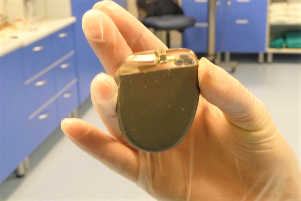
A pacemaker basically consists of 3 parts:
- A battery;
- A computerised pulse generator. Battery and pulse generator are enclosed within a small metal container, somewhat larger than the size of a two euro coin;
- One or more small cables with sensors (electrodes) at one end, called leads.
The pulse generator is the source of the electrical impulses that normalise the altered heart rhythm; the leads, on the other hand, are the connections that link the generator to the heart and allow the electrical impulses to be transmitted to the heart muscle.
The pacemaker generator is implanted under the skin
In children weighing more than 20 kg, the implantation of the generator takes place in the thoracic area, under the clavicle, with the leads going to stimulate the internal surface of the heart cavities (endocardial implantation) passing through the large veins: the subclavian vein and the superior vena cava to reach the right atrium and then the right ventricle of the heart.
In children weighing less than 15-20 kg and in those in whom it is not possible to reach the cardiac chambers from the veins, the implantation is instead cardiac surgery with the placement of the leads on the external surface of the heart (epicardial implantation) and the generator placed in a subcutaneous pocket at the level of the abdomen.
Once the implantation of the leads and the metal container has been completed, and their connection made, the pacemaker must be programmed.
Programming is carried out using a special computerised instrument and depends on the heart problem the patient is suffering from.
After setting, the pulse generator must be checked periodically to ensure that it is working properly.
Implanting a pacemaker is a fairly safe operation
However, like any surgery, it can have immediate complications such as:
- Infections at the site where the pacemaker is inserted;
- Allergic reactions to the anaesthetic drugs used during the procedure;
- Damage to blood vessels, crossed by the leads, or to nerves located near the pacemaker;
- Pulmonary collapse from haemorrhage or infiltration of air between the pleural leaflets lining the lung;
- Perforation of the myocardium;
- Swelling, haematomas and haemorrhages at the level of the pacemaker pocket.
Follow-up of the paediatric patient after surgery
The pacemaker should be checked by the doctors and technicians on a regular basis (about every 6 months), as this can happen over time:

- The cables may move or break;
- The heart condition may worsen;
- The battery may become discharged or malfunction.
Pacemaker batteries can last from 5 to 15 years (on average they last 6 or 7 years), depending on the activity of the device.
The doctor must replace the generator and the battery before the latter starts to wear out.
Some functions, including the battery status however, can also be controlled remotely via telemedicine.
It is also necessary to take a chest X-ray every 2 years to check the position and degree of tension of the leads, which may increase as the patient grows.
Read Also
Emergency Live Even More…Live: Download The New Free App Of Your Newspaper For IOS And Android
Cardiac Arrest: Why Is Airway Management Important During CPR?
RSV (Respiratory Syncytial Virus) Surge Serves As Reminder For Proper Airway Management In Children
Supplemental Oxygen: Cylinders And Ventilation Supports In The USA
What Is The Difference Between Pacemaker And Subcutaneous Defibrillator?
Heart Disease: What Is Cardiomyopathy?
Inflammations Of The Heart: Myocarditis, Infective Endocarditis And Pericarditis
Heart Murmurs: What It Is And When To Be Concerned
Broken Heart Syndrome Is On The Rise: We Know Takotsubo Cardiomyopathy
Cardiomyopathies: What They Are And What Are The Treatments
Alcoholic And Arrhythmogenic Right Ventricular Cardiomyopathy
Difference Between Spontaneous, Electrical And Pharmacological Cardioversion
What Is Takotsubo Cardiomyopathy (Broken Heart Syndrome)?
Dilated Cardiomyopathy: What It Is, What Causes It And How It Is Treated
Heart Pacemaker: How Does It Work?