
Heart pacemaker: how does it work?
A pacemaker is a cardiac stimulator consisting of a battery/generator and an electronic circuit capable of modifying the heart rate
Why is an artificial pacemaker implanted?
As mentioned in the introduction, our heartbeat is regulated by the sinoatrial node (natural pacemaker) located in the right atrium.
Under normal conditions, the heart rate is 60-80 b/min; at this rate, the heart pumps about 5 litres of blood/min.
QUALITY AED? VISIT THE ZOLL BOOTH AT EMERGENCY EXPO
Certain diseases cause an excessive slowing of the heartbeat, a condition called bradycardia, making the amount of blood and oxygen pumped by the heart to the body inadequate.
A person suffering from bradycardia may easily feel fatigued, weak, dizzy or faint.
Even normal daily activities can be tiring.
Problems may affect the natural cardiac pacemaker (SA node), which does not send stimuli at a sufficient frequency, leading to a reduction in the number of heart contractions (a slowed heartbeat is usually less than 60 b/min).
This disease is known as ‘Sick Sinus Syndrome’ or sinus node disease
Problems can also occur along the electrical stimulus conduction pathway between the atria and ventricles, electrical signals can be delayed in the AV node or fail to reach the ventricles all at once.
This condition is referred to as heart block or atrio-ventricular (AV) block.
Bradycardia can affect both very young and very old people, however it is usually diagnosed in elderly individuals.
The electrocardiographic (ECG) examination is normally used for diagnosis, sometimes additional examinations such as a dynamic ECG according to Holter with 24-hour recording or electrophysiological study are necessary.
Bradycardia in most cases is treated with the implantation of a pacemaker, which delivers electrical stimuli very similar to natural ones and modifies the heart rate according to the body’s needs.
Depending on requirements, a pacemaker can:
- replace signals from the SA node
- assist in maintaining a normal timing sequence between the upper and lower parts of the heart (atria and ventricles);
- ensure that the ventricles always contract at an appropriate frequency.
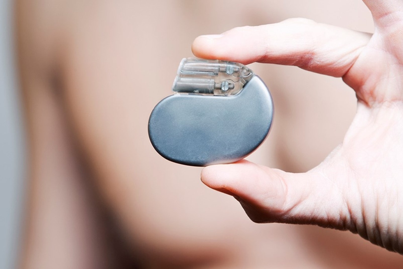
What does a pacemaker look like and how does it work?
All artificial stimulation systems (pacemakers) consist of two parts:
- the pacemaker housing the battery (about 5 cm wide, thickness
- the lead or leads, which carry impulses to the heart and transmit signals from the heart to the device.
By interpreting these signals, the pacemaker is able to monitor the heart’s activity and respond appropriately.
Modern pacemakers work ‘on demand’, i.e. they remain inactive until the natural frequency is below the set frequency.
Certain pacemaker pacing and monitoring functions can be optimally programmed or adjusted during the regularly scheduled check-ups after a pacemaker implant.
There are different types of pacemakers, mono- and bi-cameral.
Single-chamber pacemaker
The single-chamber pacemaker usually has a lead to convey signals from or to one cardiac chamber, the right atrium or more commonly the right ventricle.
This type of pacemaker is often chosen for patients in whom the SA node sends signals too slowly but whose electrical pathway to the ventricles is in good condition; for this type of patient the lead is placed in the right atrium.
Or if the SA node is functioning but the conduction system is partially or completely blocked, the lead is placed in the right ventricle.
Dual-chamber pacemaker
The dual-chamber pacemaker usually has two leads: one ends in the right atrium and the other in the right ventricle.
This type of pacemaker is capable of “feeling” (sensing function) and/or stimulating both cardiac chambers (atrium and ventricle) even separately.
The choice of a dual-chamber device can be made for several reasons.
Bi-ventricular pacemaker
In the case of a bi-ventricular pacemaker, there are three leads and they are placed in the right atrium, in the right ventricle and near the outer surface of the lateral wall of the left ventricle.
This type of pacing actually has a different indication than bradycardia and is used as supportive treatment in advanced heart failure to maintain synchronisation of the two ventricular chambers.
Some patients benefit from the implantation of a pacemaker capable of adjusting the pacing frequency to the body’s metabolic needs.
Such pacemakers are referred to as ‘frequency-modulated’ or ‘frequency-adaptive’.
In these cases, the systems use sensors that record physical parameters (such as temperature or certain body movements) to quantify the body’s metabolic needs.

How is the pacemaker implantation performed?
The pacemaker implantation procedure is performed during a surgical procedure under local anaesthesia lasting one to two hours.
The stimulator is usually implanted below the left clavicle just under the skin.
The leads are inserted into the heart through a vein located next to the collarbone, the tip of the lead is placed in contact with the endocardial tissue (inside of the heart).
Much more rarely the stimulator is placed in the abdomen and the leads are connected to the epicardium (outside of the heart), this type of procedure is performed under general anaesthesia.
After placement, the pacing system is checked. The implantation of a pacemaker usually requires a short hospitalisation (2 to 3 days).
After implantation of a pacemaker: what happens?
Most patients do not change their lifestyle (work, leisure and recreation) after pacemaker implantation.
Before being discharged from hospital, the patient receives a card that he must always carry with him, as it contains the technical specifications and programming features of the pacemaker he is carrying.
Pacemaker patients must take care to avoid activities that involve the possibility of trauma in the region of the subcutaneous pocket in which the generator is housed.
In the period immediately following implantation, the wound must be checked
It is extremely important that you follow your doctor’s instructions regarding check-ups, during which the remaining battery charge will be checked in addition to checking the function of the system.
The pacemaker is equipped with a replacement indicator that allows the physician to schedule the replacement period.
The replacement procedure is simple, usually the skin pocket is opened, the leads are disconnected (checked), connected to the new pacemaker, and the pocket is closed again.
A pacemaker is an electronic device, although it is shielded against electrical interference normally encountered, some sources can temporarily reduce or accelerate its speed.
Most household appliances and devices such as PCs, fax machines, printers are safe and do not affect the functioning of the pacemaker.
Listed below are some devices from which one should keep away or which require precautions.
Generally, these devices only temporarily affect the functioning of the pacemaker.
Transmission antennas and their power sources, avoid approaching amplifiers and linear power antennas.
Properly functioning CB radios do not cause problems.
Diathermy devices, should never be used on pacemaker patients.
Power transmission lines. Avoid high-voltage electric fields.
Electrical devices. Avoid arc welders.
Radiation. High-energy radiation can damage pacemakers. If it is necessary to undergo radiotherapy, request that lead protection be placed over the implant site.
Anti-theft-security devices Avoid standing next to anti-theft devices placed at the entrances of large stores, they can be passed at a normal pace.
Mobile telephones In some cases, mobile telephones may affect the functioning of the pacemaker if placed at a distance of less than 15 cm.
Read Also:
Emergency Live Even More…Live: Download The New Free App Of Your Newspaper For IOS And Android
What Is The Difference Between Pacemaker And Subcutaneous Defibrillator?
Heart Disease: What Is Cardiomyopathy?
Inflammations Of The Heart: Myocarditis, Infective Endocarditis And Pericarditis
Heart Murmurs: What It Is And When To Be Concerned
Broken Heart Syndrome Is On The Rise: We Know Takotsubo Cardiomyopathy
Cardiomyopathies: What They Are And What Are The Treatments
Alcoholic And Arrhythmogenic Right Ventricular Cardiomyopathy
Difference Between Spontaneous, Electrical And Pharmacological Cardioversion
What Is Takotsubo Cardiomyopathy (Broken Heart Syndrome)?
Dilated Cardiomyopathy: What It Is, What Causes It And How It Is Treated