
Circulatory assistance to the left ventricle: the intra-aortic counterpulsation
The intra-aortic counterpulsation is a device that is used in cardiology because it is capable of providing temporary circulatory assistance
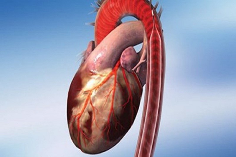
It is a mechanical support for the left ventricle of the heart, the cavity that pumps blood to the aorta.
Its operation decreases myocardial oxygen demand by increasing cardiac output with the effect of increasing coronary blood flow and oxygen supply.
The device was pioneered at Grace Sinai Hospital in Detroit in the 1960s by Dr. Kantrowitz and his team.
The first clinical implantation was conducted at Maimonides Medical Center in Brooklyn in October 1967, on a 48-year-old woman in cardiogenic shock who was not responding to conventional therapy.
The intra-aortic counterpulsation was inserted with a downward cut of the left femoral artery
The pumping was performed for about 6 hours, the state of shock resolved and the patient was discharged.
The device was developed for use in cardiac surgery by Dr. David Bregman in 1976 at New York-Presbyterian Hospital in New York.
In 1978, Dr. Subramanian experimented with insertion using the Seldinger technique, i.e. with percutaneous access, which facilitated its use.
The myocardial cells are perfused with oxygenated blood, inflating the balloon in the thoracic aorta at the time of greatest coronary artery filling, diastole, so as to increase the systolic blood pressure by supporting the function of the left ventricle by decreasing peripheral resistance.
In systole, the rapidly deflating balloon creates a reduction in cardiac afterload resulting in a reduction in myocardial oxygen consumption and an increase in cardiac output.
Composition and function of the intraortic counterpulsation
The intra-aortic counterpulsation is a system consisting of an external mechanical part and a catheter with a balloon that is introduced percutaneously into the patient’s thoracic aorta via the femoral artery, through a procedure performed under local anaesthesia and supported by X-rays.
The aortic counterpulsator consists of a semi-rigid vascular catheter on the distal portion of which is mounted a polyethylene balloon connected via a tube to the machine body (console) capable of synchronising the insufflation and deflation of the balloon to the cardiac cycle.
The sterile disposable kit containing the catheter consists of two separate trays, the first contains all the material necessary to position the percutaneous arterial access, the second contains the catheter with balloon accompanied by tubing and cables to be connected to the machine body.
The counterpulsation consists of a pneumatic part and an electronic part; the pneumatic/mechanical part is connected to the balloon, which allows it to be inflated and deflated with each cardiac cycle.
The electronic part, which regulates and controls, synchronises and monitors the operation of the entire system.
The counterpulsation can be synchronised with the pressure wave or with the electrocardiographic trace, by means of 5 electrodes applied to the patient’s chest.
The physician then optimises the timing of inflation and deflation of the balloon and adjusts the service ratio.
The monitor displays ECG, pressure curve and inflation/deflation cycles, highlighting the pressures measured in real time.
The control unit operates the pneumatic system, which uses helium (inert gas) contained in a cylinder inside the console, to inflate and deflate the balloon placed in the aorta.
The balloon is dilated in diastole and deflated in systole.
The device decreases the workload on the heart, allowing it to pump more blood.
When the left ventricle has finished pumping blood, diastole, the device dilates: this will increase blood flow to the heart and the rest of the body.
When the left ventricle is about to pump blood, systole, the balloon deflates: this creates extra space in the aorta allowing the heart to pump more blood.
The aortic counterpulsator catheter has a variable size, which is selected according to the patient’s build; the substance with which the balloon is inflated is helium, an inert gas whose chemical/physical properties prevent it from creating embolisms in the event of a rupture.
To position it, after disinfecting the groin, the femoral artery is punctured and the introducer is placed.
The catheter is gently removed from the tray, and while it is still in the pack, the connectors are passed to the theatre nurse who will insert the calibration key and the fibre optic connector into the body.
Next, the spindle is removed from the lumen of the counterpulsator catheter and flushed with heparin saline, then a one-way valve is placed over the lumen connected to the balloon and a vacuum is created using a syringe.
The haemodynamist can introduce the catheter by sliding it over the metal guide wire; the catheter must be positioned precisely, its tip must reach just below the branch of the left subclavian artery, while the distal end must be above the emergence of the renal arteries.
Once the correct positioning has been checked by fluoroscopy, the one-way valve is removed from the lumen of the balloon and the helium tube is connected; counterpulsation can begin.
While the haemodynamist secures the catheter with stitches in the thigh, the theatre nurse connects the ECG leads to the patient so that the counterpulsation can be activated even if the fibre-optic pressure signal is disturbed.
Finally, a pressure infusion of heparin saline is connected to the lumen of the catheter.
Since the device is inserted into the femoral artery and aorta, it may cause tissue ischaemia.
The leg is at higher risk of being affected by ischaemia if the femoral artery is obstructed.

Placement of the balloon too distal from the aortic arch may induce occlusion of the renal artery resulting in renal failure.
Other possible complications are cerebral embolism during insertion, infection, dissection of the aorta or iliac artery, perforation of the artery and subsequent bleeding in the mediastinum.
Any mechanical failure of the balloon may require emergency vascular surgery to remove it.
The haemodynamics nurse has a broad responsibility for the management of the aortic counterpulsator: he/she must be able to provide routine maintenance of the machine to ensure its proper functioning.
It is the responsibility of the nurse to scrupulously supervise the patient in order to avoid the occurrence of possible life-threatening complications, such as haemorrhage, displacement of the counterpulsation catheter and arrhythmias.
Read Also
Emergency Live Even More…Live: Download The New Free App Of Your Newspaper For IOS And Android
Myocardiopathy: What Is It And How To Treat It?
Venous Thrombosis: From Symptoms To New Drugs
Cyanogenic Congenital Heart Disease: Transposition Of The Great Arteries
Heart Murmur: What Is It And What Are The Symptoms?
Branch Block: The Causes And Consequences To Take Into Account
Cardiopulmonary Resuscitation Manoeuvres: Management Of The LUCAS Chest Compressor
Supraventricular Tachycardia: Definition, Diagnosis, Treatment, And Prognosis
Identifying Tachycardias: What It Is, What It Causes And How To Intervene On A Tachycardia
Myocardial Infarction: Causes, Symptoms, Diagnosis And Treatment
Aortic Insufficiency: Causes, Symptoms, Diagnosis And Treatment Of Aortic Regurgitation
Congenital Heart Disease: What Is Aortic Bicuspidia?
Atrial Fibrillation: Definition, Causes, Symptoms, Diagnosis And Treatment
Ventricular Fibrillation Is One Of The Most Serious Cardiac Arrhythmias: Let’s Find Out About It
Atrial Flutter: Definition, Causes, Symptoms, Diagnosis And Treatment
What Is Echocolordoppler Of The Supra-Aortic Trunks (Carotids)?
What Is The Loop Recorder? Discovering Home Telemetry
Cardiac Holter, The Characteristics Of The 24-Hour Electrocardiogram
Peripheral Arteriopathy: Symptoms And Diagnosis
Endocavitary Electrophysiological Study: What Does This Examination Consist Of?
Cardiac Catheterisation, What Is This Examination?
Echo Doppler: What It Is And What It Is For
Transesophageal Echocardiogram: What Does It Consist Of?
Paediatric Echocardiogram: Definition And Use
Heart Diseases And Alarm Bells: Angina Pectoris
Fakes That Are Close To Our Hearts: Heart Disease And False Myths
Sleep Apnoea And Cardiovascular Disease: Correlation Between Sleep And Heart