
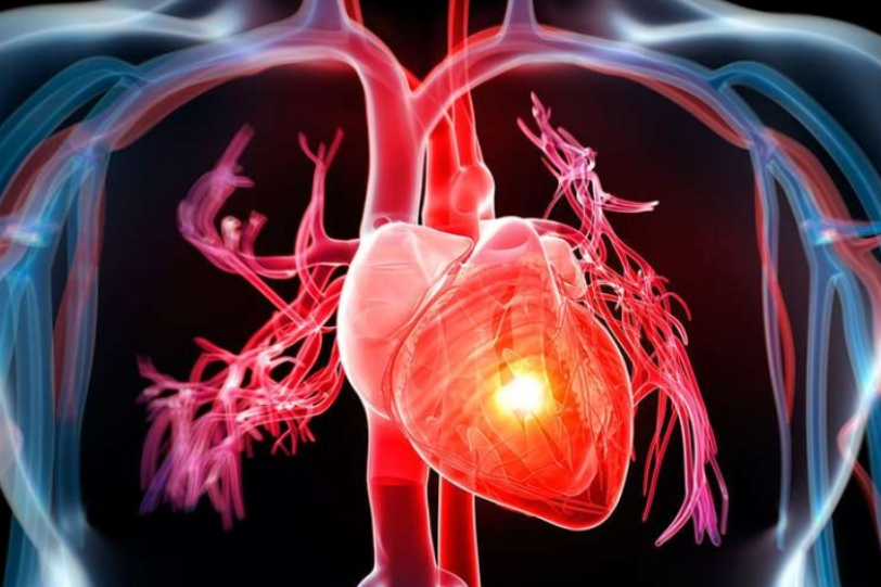
Heart diseases and alarm bells: angina pectoris
Angina pectoris is a heart condition that is an important alarm bell for possible coronary artery disease
Angina pectoris: what is it?
Angina pectoris is a term derived from the Latin words angina=pain and pectoris=pain.
It can be of three types:
- Stable or exertional angina: generally arises during physical exertion, but also in cold weather or during emotional stress and in general in all those situations that require an increased blood flow to the heart;
- Unstable angina: pain occurs suddenly, even at rest, or during physical exertion that is not particularly demanding. It may be caused by a temporary obstruction of a coronary artery (e.g. due to a thrombus or the formation of fibrous plaques along the vessel walls) or by a spasm of the same.
- Secondary angina: refers to all forms of cardiac ‘ischaemia’ not caused by coronary narrowing or obstruction, but secondary to other pathologies such as mitral and aortic valvulopathy, severe anaemia, hyperthyroidism and arrhythmias.
What are the causes of angina pectoris?
Angina pectoris can be caused by a temporary poor blood supply to the coronary arteries, i.e. the arteries of the heart, resulting in a lack of oxygen to the heart muscle.
The mechanism that causes the narrowing or even complete obstruction of the coronary arteries is atherosclerosis, which is caused by intravascular deposits of fat (cholesterol and triglycerides).
What are the symptoms of angina pectoris?
The main symptom of angina is chest pain, which can be classified according to the following parameters:
- Quality: oppressive, constrictive, sharp, dull, varying in intensity from mild to severe;
- Localisation: typically referred to the retrosternal region; may in some cases affect the whole chest and radiate to the neck, jaw, arms, wrists and shoulders
- Duration: a few minutes or longer
- Frequency: sporadic, regular, irregular, frequent episodes.
In addition to pain, angina pectoris may be accompanied by:
- nausea
- tiredness
- dizziness
- difficulty breathing
- restlessness
What are the risk factors for angina pectoris?
The causes of angina pectoris can be found in all those risk factors that cause damage to the walls of the coronary arteries, such as
- smoking, alcohol, drugs
- high blood pressure;
- diabetes mellitus;
- dyslipidemia;
- obesity;
- sedentariness;
- early family history of cardiovascular disease;
- diet with excess calories, salt, saturated fat, simple sugars and cholesterol; diet low in fibre, vitamins, fish and polyunsaturated fatty acids.
Angina pectoris, what tests should be done for diagnosis?
The diagnosis of angina is made by the doctor first of all on the basis of the data obtained from the interview with the patient (anamnesis).
In addition, several tests can be performed including:
- Electrocardiogram (ECG): test that records the electrical activity of the heart. It can detect any abnormalities or alterations in the heart rhythm using, for example, the dynamic electrocardiogram according to Holter, recording the electrocardiographic tracing over a prolonged period (usually 24 hours);
- Colour Doppler echocardiogram: a test that uses ultrasound to obtain a morphological and functional assessment of the heart. It is a method that makes it possible to study the contractility of the heart, the morphology of the valves and the flow of blood in its cavities, both at rest and after exercise or after taking a drug;
- Exercise test: a test that involves observing the heart’s response to exercise. It is usually performed on a treadmill or exercise bike, while the patient is monitored by ECG;
- Computed tomography of the heart (CT heart scan): allows visualisation, using contrast medium, of the anatomy of the coronary arteries and early detection and quantification of any occlusions or narrowings (stenosis) and atherosclerotic plaques that may cause significant reductions in blood flow to the heart muscle;
- Stress Magnetic Resonance Imaging (MRI) is an MRI test aimed at assessing the efficiency of the coronary circulation, the presence of previous heart attacks and the functioning of the heart under stress conditions;
- Coronary angiography (coronarography): a test that involves inserting a catheter through an artery in the arm or leg into the coronary arteries of the heart. A contrast agent is then injected, which makes it possible to visualise any blockages or stenosis of the coronary arteries.
The specialist may decide to perform one or more of these tests, depending on the severity of the symptoms and the patient’s condition.
In any case, early diagnosis of angina pectoris is essential to prevent more serious complications and to ensure proper management of cardiac health.
How angina pectoris is treated
Treatment for stable angina pectoris can include several treatment options.
Lifestyle changes and reversible risk factors: changes in diet, regular exercise, quitting smoking and reducing stress, correcting high blood pressure, diabetes mellitus and dyslipidaemia may be recommended
Drug therapy, different types of drugs are prescribed including
- Beta-blockers, which reduce myocardial oxygen demands and increase myocardial exercise tolerance.
- Calcium channel blockers, which with different mechanisms can be used for hypertension or coronary spasm.
- Inhibitors of angiotensin-converting enzyme (ACEi), and angiotensin AT1 receptor antagonists (sartans), which are used for the treatment of hypertension and for post ischaemic heart remodelling.
- Statins, drugs to control cholesterol that limit its production and accumulation on artery walls, slowing the development or progression of atherosclerosis.
- Antiplatelet drugs (aspirin, clopidogrel, prasugrel or ticagrelor) to inhibit platelet aggregation and prevent thrombus formation.
- Other categories of drugs are used to treat angina, improve coronary artery perfusion and prevent the risk of heart attack and thrombosis.
Interventional medical procedures

- Procedures such as percutaneous coronary angioplasty may be recommended, which involves inserting a small balloon usually associated with a metal mesh structure (stent) into the lumen of the coronary artery, which is inflated at the narrowing of the obstructed artery, improving blood flow downstream
- Coronary artery bypass surgery, which involves creating a new pathway for blood flow through the use of a patient’s own artery or vein to ‘bypass’ the point of narrowing of the coronary artery, thereby causing the upstream portion to communicate directly with the downstream portion of the stenosis.
Therapy for angina pectoris must be customised for each patient and depends on the severity of the symptoms and the patient’s health condition.
It is important to carefully follow the doctor’s instructions and adopt a healthy lifestyle to prevent complications and improve quality of life.
Read Also
Emergency Live Even More…Live: Download The New Free App Of Your Newspaper For IOS And Android
Myocardiopathy: What Is It And How To Treat It?
Venous Thrombosis: From Symptoms To New Drugs
Heart Murmur: What Is It And What Are The Symptoms?
Cardiopulmonary Resuscitation Manoeuvres: Management Of The LUCAS Chest Compressor
Supraventricular Tachycardia: Definition, Diagnosis, Treatment, And Prognosis
Identifying Tachycardias: What It Is, What It Causes And How To Intervene On A Tachycardia
Myocardial Infarction: Causes, Symptoms, Diagnosis And Treatment
Aortic Insufficiency: Causes, Symptoms, Diagnosis And Treatment Of Aortic Regurgitation
Congenital Heart Disease: What Is Aortic Bicuspidia?
Atrial Fibrillation: Definition, Causes, Symptoms, Diagnosis And Treatment
Ventricular Fibrillation Is One Of The Most Serious Cardiac Arrhythmias: Let’s Find Out About It
Atrial Flutter: Definition, Causes, Symptoms, Diagnosis And Treatment
What Is Echocolordoppler Of The Supra-Aortic Trunks (Carotids)?
What Is The Loop Recorder? Discovering Home Telemetry
Cardiac Holter, The Characteristics Of The 24-Hour Electrocardiogram
Peripheral Arteriopathy: Symptoms And Diagnosis
Endocavitary Electrophysiological Study: What Does This Examination Consist Of?
Cardiac Catheterisation, What Is This Examination?
Echo Doppler: What It Is And What It Is For
Transesophageal Echocardiogram: What Does It Consist Of?
Paediatric Echocardiogram: Definition And Use