

Spinal shock: causes, symptoms, risks, diagnosis, treatment, prognosis, death
Spinal distribution shock: ‘shock’ in medicine refers to a syndrome, i.e. a set of symptoms and signs, caused by reduced perfusion at a systemic level with an imbalance between oxygen availability and its demand at a tissue level
Shock is classified into two major groups
- decreased cardiac output shock: cardiogenic, obstructive, haemorrhagic hypovolaemic and non-haemorrhagic hypovolaemic;
- distributive shock (from decreased total peripheral resistance): septic, allergic (‘anaphylactic shock’), neurogenic and spinal.
Spinal distributive shock
Distributive shock is a type of shock caused by a disproportion between the vascular bed, which is abnormally dilated, and the circulating blood volume, which – although not absolutely reduced – becomes insufficient due to the vasodilation created.
Spinal shock is a rare type of distributive shock in which peripheral vasodilation is caused by injury to the spinal cord contained in the spinal column.
This form should not be confused with a similar one, neurogenic shock.
In several texts, the two types of shock are associated, but in the case of spinal shock, a loss of spinal cord-mediated reflexes is observed.
Shock is often the first manifestation of a spinal cord injury.
TRAINING IN FIRST AID? VISIT THE DMC DINAS MEDICAL CONSULTANTS BOOTH AT EMERGENCY EXPO
In this type of spinal shock there is, to simplify, this sequence of events:
- nerve damage results in a decrease in the nerve mechanisms controlling the blood circulation;
- peripheral vasodilatation occurs;
- peripheral vasodilatation leads to arterial hypotension;
- arterial hypotension leads to tissue hypoperfusion;
- tissue hypoperfusion leads to tissue anoxia;
- ischaemic distress leads to necrosis (death) of tissues, which stop functioning.
Symptoms and signs of spinal shock
The following clinical signs and symptoms can be seen in this type of shock:
- arterial hypotension
- fatigue;
- altered respiratory rate;
- bradycardia or tachycardia (decreased or increased heart rate);
- symptoms and signs of multi-organ dysfunction;
- collapse of blood pressure;
- cardiac arrest;
- pulmonary arrest;
- severe reduction in level of consciousness;
- coma;
- death.
These symptoms and signs must also be associated with other symptoms and signs caused by the upstream condition and/or pathology that caused the shock, such as those of spinal cord compression, which can lead to motor deficits (e.g. paralysis of the lower limbs or even the upper limbs in the case of cervical vertebra injury) and sensory deficits.
Loss of sensation and movement occurs below the site of injury, so the higher the injury (e.g. cervical vertebra fracture), the more severe the damage will generally be.
RESCUE RADIO IN THE WORLD? VISIT THE EMS RADIO BOOTH AT EMERGENCY EXPO
Other immediate symptoms may include:
- pain in the area of the injury
- muscle spasticity;
- tingling and numbness in the limbs;
- priapism in males;
- dyspnoea;
- respiratory failure;
- cardiac arrhythmia;
- loss of bladder function;
- loss of bowel function.
Long-term effects of spinal trauma vary depending on the location and severity of the injury: as already mentioned, the higher the damage in the spine, the more severe, in general, are the symptoms.
For example, an injury to the cervical spine will affect all four limbs, as well as the muscles that control breathing and other essential functions.
An injury to the lumbar spine, on the other hand, will affect the lower limbs (not the upper limbs) and the bowel and bladder function, but usually does not affect other organs or systems.
Complete high neck injury and trauma complicated by other serious injuries may cause immediate death or result in severe impairment of autonomy, eventually requiring total assistance for the rest of the patient’s life.
Stages of spinal shock
This type of shock is distinguished into four different phases based on the course of the reflexes:
- phase 1 loss of reflexes (areflexia);
- phase 2 after about two days part of the reflexes are recovered;
- phase 3 hyperreflexia occurs;
- phase 4 spastic phase.
According to other authors, spinal shock can be divided into two phases:
– acute phase
- areflexia;
- retention of evacuation routes;
- vasoparalysis;
- skin hypothermia;
- paraplegia;
- muscular hypotonia;
– chronic phase:
- hyperflexia;
- spasticism;
- spinal automatism.
These phases generally comprise a time span of three to six weeks; in some cases the total duration of these phases has been several months.
In the period immediately following the injury (lasting hours or days), spinal shock is characterised by flaccidity, loss of autonomic functions and complete anaesthesia below the injury, which last the longer the injury itself is in the upper part of the spine; this picture is progressively succeeded by spasticity.
Causes and risk factors of spinal shock
Pathologies and conditions that most frequently cause and/or promote neurogenic shock are spinal cord injuries with quadriplegia or paraplegia.
A frequent trauma is the fracture of a vertebra and/or its dislocation, resulting in compression and/or injury of the spinal cord.
Such types of trauma often occur in traffic or sports accidents, or in falls or injuries caused by gunshots.
Spinal cord trauma can be
- direct (closed or penetrating);
- related to exceeding the limits of movement granted to the spinal cord within the spinal canal (excessive hyperextension, hyperflexion or torsion).
Spinal shock is also sometimes a consequence of spinal tumours or an abnormality that may occur after birth due to stress-related events.
Course of spinal shock
Three different phases can generally be identified in a shock:
- initial compensatory phase: the cardiovascular depression worsens and the body triggers compensation mechanisms mediated by the sympathetic nervous system, catecholamines and production of local factors such as cytokines. The initial phase is more easily treatable. Early diagnosis leads to a better prognosis, however it is often arduous as symptoms and signs may be blurred or non-specific at this stage;
- progression phase: the compensation mechanisms become ineffective and the perfusion deficit to vital organs worsens rapidly, causing severe pathophysiological imbalances with ischaemia, cellular damage and accumulation of vasoactive substances. Vasodilation with increased tissue permeability can lead to disseminated intravascular coagulation.
- irreversibility phase: this is the most severe phase, where marked symptoms and signs facilitate diagnosis, which, however, performed at this stage, often leads to ineffective therapies and a poor prognosis. Irreversible coma and reduced cardiac function may occur, up to cardiac arrest and death of the patient.
Diagnosis of spinal shock
The diagnosis of shock is based on various tools, including:
- anamnesis;
- objective examination;
- laboratory tests;
- haemochrome;
- haemogasanalysis;
- CT SCAN;
- coronarography;
- pulmonary angiography;
- electrocardiogram;
- chest X-ray;
- echocardiogram with colordoppler.
The most common examinations used for the differential diagnosis are CT scan, echocardiography, cardiac catheterisation, abdominal ultrasound, as well as laboratory tests to rule out haemorrhages and coagulation disorders.
Anamnesis and objective examination are important and must be performed very quickly.
In the case of an unconscious patient, the history can be taken with the help of family members or friends, if present.
On objective examination, the subject with shock often presents pale, with cold, clammy skin, tachycardic, with reduced carotid pulse, impaired renal function (oliguria) and impaired consciousness.
During diagnosis, it will be necessary to ensure airway patency in patients with impaired consciousness, place the subject in the anti-shock position (supine), cover the casualty, without making him sweat, to prevent lipotimia and thus further aggravation of the state of shock.
With regard to laboratory tests, fundamental in the diagnosis of shock is arterial or venous haemogasanalysis, to assess the body’s acid-base balance.
Characteristically, shock is accompanied by a picture of metabolic acidemia with increased lactates and base deficiency.

CT and MRI scans of the spine are essential to detect spinal cord damage
Diagnosis and management of spinal cord injury can be difficult and injuries that are not diagnosed early can cause serious complications.
If a spinal cord injury is suspected, the spine must be protected and immobilised at all times during evaluation and diagnosis.
The initial assessment includes medical history, clinical examination and above all imaging (X-ray, CT scan, MRI), which must include the entire spine, not just the region where the injury is suspected.
The choice of diagnostic techniques varies depending on the patient’s state of consciousness and the presence of other injuries.
In spinal distribution shock, this situation occurs:
- preload: decreases/normal
- afterload: decreases;
- contractility: normal;
- central venous satO2: varies; in arteriovenous shunt there is an increase;
- Hb concentration: normal;
- diuresis: normal/decreased;
- peripheral resistance: decreased;
- sensory: normal in neurogenic and spinal shock; agitation/confusion in septic and allergic shock.
Let us remember that systolic output depends by Starling’s law on preload, afterload and contractility of the heart, which can be monitored clinically indirectly by various methods:
- preload: by measuring the central venous pressure through the use of the Swan-Ganz catheter, bearing in mind that this variable is not in linear function with preload, but this also depends on the rigidity of the walls of the right ventricle;
- afterload: by measuring systemic arterial pressure (in particular diastolic, i.e. the ‘minimum’);
- contractility: by echocardiogram or myocardial scintigraphy.
The other important parameters in the case of shock are checked by:
- haemoglobin: by haemochrome;
- oxygen saturation: by means of a saturation meter for the systemic value and by taking a special sample from the central venous catheter for venous saturation (the difference with the arterial value indicates oxygen consumption by the tissues)
- arterial oxygen pressure: via haemogasanalysis
- diuresis: via bladder catheter.
During diagnosis, the patient is observed continuously, to check how the situation evolves, always keeping the ‘ABC rule’ in mind, i.e. checking
- patency of the airways
- presence of breathing;
- presence of circulation.
These three factors are vital for the patient’s survival, and must be controlled – and if necessary re-established – in that order.
Therapy
Therapy depends on the upstream cause of shock. Oxygen administration is usually carried out, followed by adjustment of the individual’s fluids to restore proper volaemia: isotonic crystalloids are used for this purpose; in more severe cases where normal therapy appears to be unsuccessful, dopamine or noradrenaline is used.
Specifically, therapy includes
- immobilisation of the head, neck and back;
- implementation of specific measures relating to the upstream cause of shock, e.g. neurological and/or orthopaedic surgical therapy in the case of tumours and/or traumatic injuries of vertebrae and spinal cord;
- withdrawal of vasodilator drugs;
- volaemia expansion: infusion of e.v. crystalloid solution (1 litre over 20-30 minutes, continuing until central venous pressure values normalise). Colloids may also be used in this type of shock;
- vasoconstrictor drugs: these counter peripheral vasodilation and arterial hypotension. The administration of dopamine in doses of 15-20 mg/kg/minute or noradrenaline in doses of 0.02-0.1 mcg/kg/minute is useful (the infusion should be adjusted so as not to exceed 100 mmHg systolic blood pressure).
Rehabilitation in spinal shock:
In addition to the therapies listed above, physiotherapeutic rehabilitation treatments are combined over time to restore as much as possible the sensory and/or motor function lost due to the spinal cord injury.
Physical, occupational, speech and rehabilitation therapy are important parts of the long-term recovery process.
Rehabilitation focuses on the prevention of muscle atrophy and contracture, helps patients learn to retrain some of their muscles to compensate for the loss of others, and can improve communication in a patient who has lost some ability to speak and move.
Unfortunately, treatments do not always yield the results the patient hopes for.
Depending on the severity of the injury, long-term interventions may be necessary to maintain everyday functions, for example they may include:
- mechanical ventilation to facilitate breathing;
- bladder catheter to drain the bladder;
- feeding tube to provide additional nutrition and calories.
Evolution and prognosis of spinal shock
Severe spinal shock that is not treated quickly often has a poor prognosis, especially in the case of cervical vertebra injury.
Even when medical intervention is timely, the prognosis is sometimes inauspicious.
Once the process triggering the syndrome has begun, tissue hypoperfusion leads to a multi-organ dysfunction, which increases and worsens the state of shock: various substances are poured into the circulatory stream from vasoconstrictors such as catecholamines, to various kinins, histamine, serotonin, prostaglandins, free radicals, complement system activation and tumour necrosis factor.
All these substances do nothing but damage vital organs such as the kidney, heart, liver, lung, intestines, pancreas and brain.
Severe spinal shock that is not treated in time has a poor prognosis, as it can lead to irreversible motor and/or sensory nerve damage, coma and death of the patient.
Lasting from a few hours to a couple of weeks, spinal shock may subside over time to reveal the true extent of the damage, which, however, is often severe and irreversible, with little response to rehabilitation therapy.
What to do?
If you suspect that someone is suffering from shock, contact the Single Emergency Number.
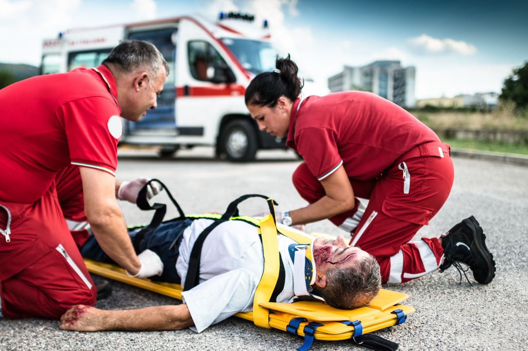
The subject is immobilised starting with the neck, which is locked with a neck brace, after which the back, upper limbs, pelvis and lower limbs are immobilised.
For this purpose, straps or belts can be used to immobilize the subject’s movements.
If possible, place the subject in the anti-shock position, or Trendelenburg position, which is achieved by placing the casualty lying on the floor, supine, tilted 20-30° with the head on the floor without a pillow, with the pelvis slightly elevated (e.g. with a pillow) and the lower limbs raised.
Read Also:
Emergency Live Even More…Live: Download The New Free App Of Your Newspaper For IOS And Android
Electrical Injuries: How To Assess Them, What To Do
RICE Treatment For Soft Tissue Injuries
How To Carry Out Primary Survey Using The DRABC In First Aid
Heimlich Maneuver: Find Out What It Is And How To Do It
What Should Be In A Paediatric First Aid Kit
Poison Mushroom Poisoning: What To Do? How Does Poisoning Manifest Itself?
Hydrocarbon Poisoning: Symptoms, Diagnosis And Treatment
First Aid: What To Do After Swallowing Or Spilling Bleach On Your Skin
Signs And Symptoms Of Shock: How And When To Intervene
Wasp Sting And Anaphylactic Shock: What To Do Before The Ambulance Arrives?
UK / Emergency Room, Paediatric Intubation: The Procedure With A Child In Serious Condition
Endotracheal Intubation In Paediatric Patients: Devices For The Supraglottic Airways
Sedation And Analgesia: Drugs To Facilitate Intubation
Intubation: Risks, Anaesthesia, Resuscitation, Throat Pain