
What is breast ultrasound?
Breast ultrasound is not a screening test. In the presence of a clinical symptom it may represent a first level test allowing to verify the origin of the symptom itself (solid or liquid lump, inflammatory collection, etc.)
Ultrasound is a diagnostic imaging test based on the use of ultrasound, therefore, noninvasive
When applied to the breast, it allows the study of the breast parenchyma and axillary cords.
In asymptomatic women, ultrasonography finds indication mainly as a second-level investigation following the performance of a mammogram and the finding of a suspicious alteration or a tissue with high density, that is, rich in fibro-ghiandular component.
Finally, ultrasonography allows exploration of the axillary cords normally poorly evaluated by mammography alone.
Who can perform breast ultrasound?
All women but with different indications according to different age groups.
Although in women younger than 40 years of age, in relation to the low incidence of breast cancer, there is no indication for preventive examinations, in the presence of significant familiarity (multiple cases of breast cancer in first- and/or second-degree relatives) it is advisable to perform breast ultrasound from the age of 30 years.
The test is not contraindicated in pregnancy but is not recommended in that condition because of increased parenchyma density in the gestation period.
When necessary, it may be suggested to characterize clinically appreciable lesions.
Ultrasonography, performed as a diagnostic adjunct to a mammogram in women older than 40 years and with dense breasts, increases their diagnostic ability by increasing their sensitivity, that is, their ability to recognize tumors.
Is breast ultrasound a painful or dangerous test?
It is an absolutely harmless and certainly not painful test, which may result in only slight discomfort in particularly sensitive patients.
How does it work?
Ultrasound is a diagnostic imaging technique based on the analysis of the echoes produced by an ultrasound beam passing through an organ or tissue.
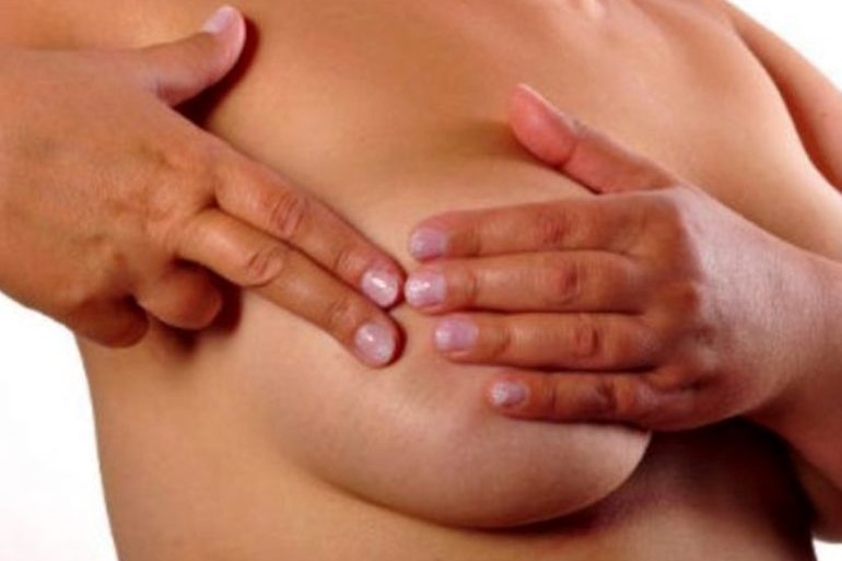
In breast ultrasound, the radiologist performs the study of both breasts and axillary cords using a high-frequency probe specifically designed for soft tissue.
The test is performed with the patient lying supine and hands clasped behind the head; a gel is applied between the probe and skin to prevent the interposed air from blocking the ultrasound.
The complete procedure takes about 15 minutes.
Read Also
Emergency Live Even More…Live: Download The New Free App Of Your Newspaper For IOS And Android

What Is The Resting Cardiac Echocolordoppler (Or Echocardiogram)?
What Is Echocardiography (Echocardiogram)?
Diagnostic Tests: What Is Ultrasound?
Echocardiogram: What It Is And When It Is Required
What Is Echocolordoppler Of The Supra-Aortic Trunks (Carotids)?
What Is The Loop Recorder? Discovering Home Telemetry
Cardiac Holter, The Characteristics Of The 24-Hour Electrocardiogram
Peripheral Arteriopathy: Symptoms And Diagnosis
Endocavitary Electrophysiological Study: What Does This Examination Consist Of?
Cardiac Catheterisation, What Is This Examination?
Echo Doppler: What It Is And What It Is For
Transesophageal Echocardiogram: What Does It Consist Of?
Venous Thrombosis: From Symptoms To New Drugs
Echotomography Of Carotid Axes
Echo- And CT-Guided Biopsy: What It Is And When It Is Needed
Echodoppler: What It Is And When To Perform It
What Is 3D Echocardiography (Echocardiogram)?
Congenital Heart Disease: Tricuspid Atresia
What Is Stress Echocardiography And What Is It Used For?